Copyright
©The Author(s) 2022.
World J Clin Cases. Apr 26, 2022; 10(12): 3834-3841
Published online Apr 26, 2022. doi: 10.12998/wjcc.v10.i12.3834
Published online Apr 26, 2022. doi: 10.12998/wjcc.v10.i12.3834
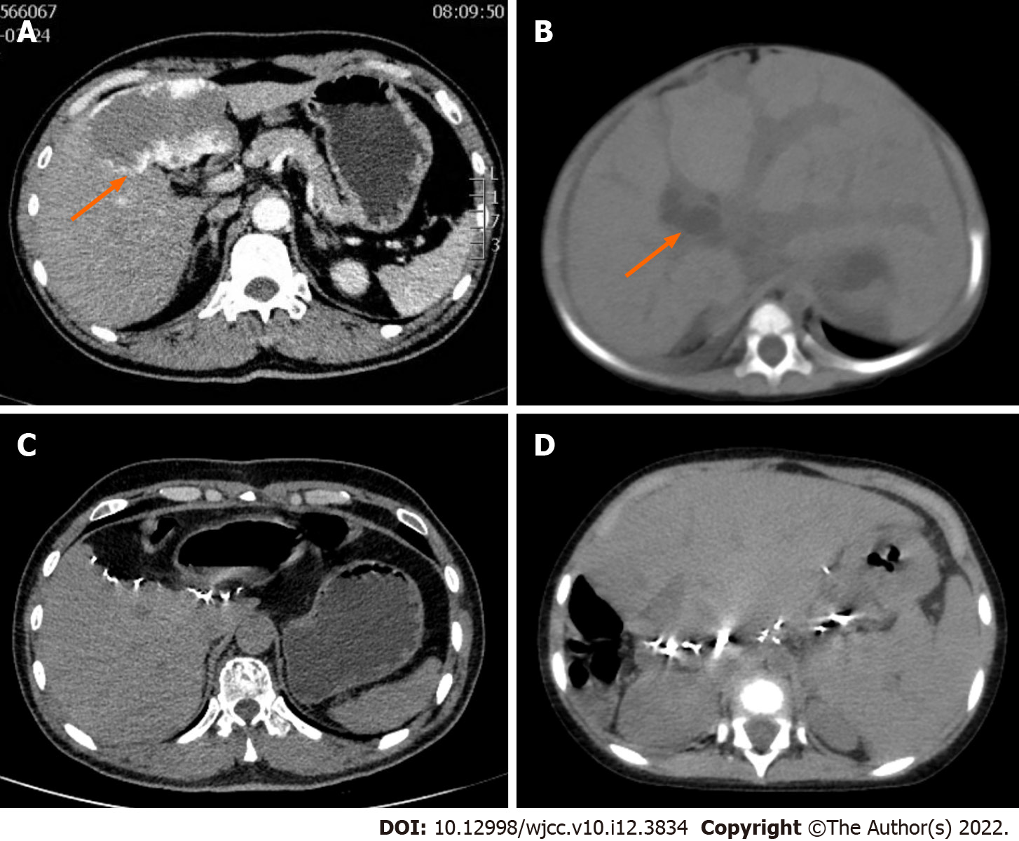
Figure 1 Preoperative contrast-enhanced abdominal computed tomography and postoperative abdominal computed tomography of the donor and recipient liver.
A: Hemangioma was indicated by orange arrow; B: Dilated intrahepatic bile duct showed by orange arrow, and diffuse hepatomegaly could be seen; C: The liver of the donor was regenerated half a year after the operation without hemangioma recurrence; D: The liver allograft transplanted to the recipient was regenerated 2 years after the operation without hemangioma recurrence.
- Citation: Li SX, Tang HN, Lv GY, Chen X. Pediatric living donor liver transplantation using liver allograft after ex vivo backtable resection of hemangioma: A case report. World J Clin Cases 2022; 10(12): 3834-3841
- URL: https://www.wjgnet.com/2307-8960/full/v10/i12/3834.htm
- DOI: https://dx.doi.org/10.12998/wjcc.v10.i12.3834


