Copyright
©The Author(s) 2022.
World J Clin Cases. Apr 26, 2022; 10(12): 3686-3697
Published online Apr 26, 2022. doi: 10.12998/wjcc.v10.i12.3686
Published online Apr 26, 2022. doi: 10.12998/wjcc.v10.i12.3686
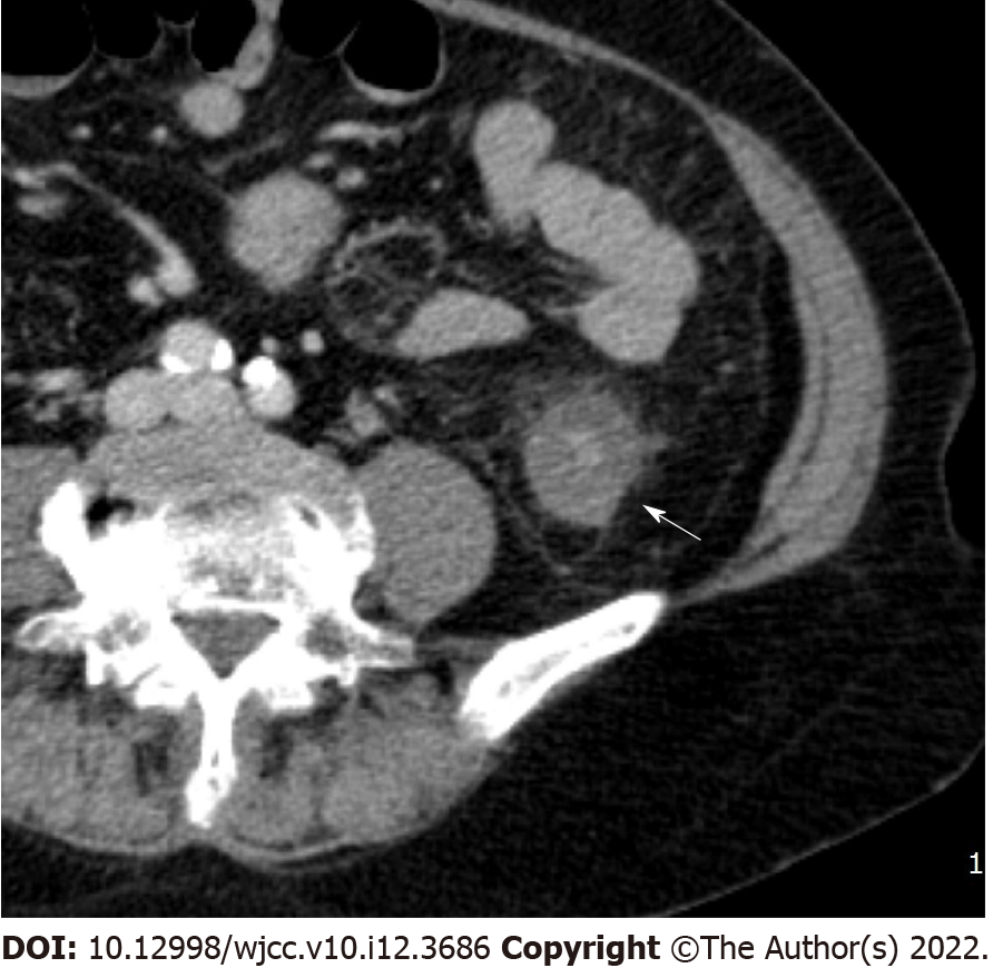
Figure 1 Serosal involvement.
Enhanced multidetector computed tomography axial image in portal venous phase shows wall thickening with submucosal edema and pericolic fat stranding (arrow) in descending colon.
- Citation: Yu SJ, Heo JH, Choi EJ, Kim JH, Lee HS, Kim SY, Lim JH. Role of multidetector computed tomography in patients with acute infectious colitis. World J Clin Cases 2022; 10(12): 3686-3697
- URL: https://www.wjgnet.com/2307-8960/full/v10/i12/3686.htm
- DOI: https://dx.doi.org/10.12998/wjcc.v10.i12.3686


