Copyright
©The Author(s) 2022.
World J Clin Cases. Apr 16, 2022; 10(11): 3630-3638
Published online Apr 16, 2022. doi: 10.12998/wjcc.v10.i11.3630
Published online Apr 16, 2022. doi: 10.12998/wjcc.v10.i11.3630
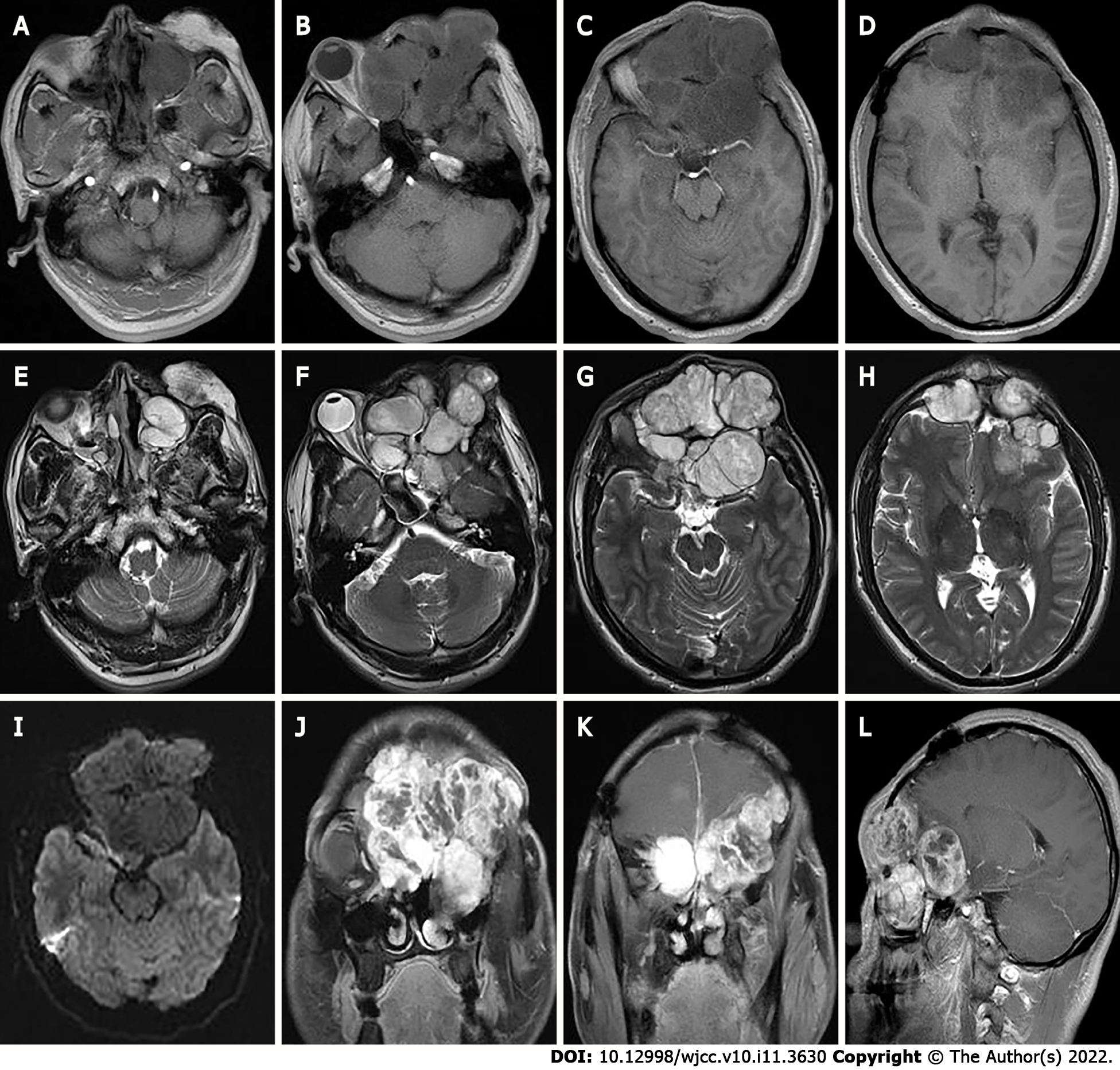
Figure 4 The second preoperative magnetic resonance imaging findings.
A-H: Magnetic resonance imaging shows multiple masses of T1 weighted imaging (WI) low signals (A-D) and T2WI inhomogeneous high signals (E-H) in the left orbit, bilateral frontal areas, bilateral septal sinuses, pterygoid sinuses, left maxillary sinus, and anterior skull base; I: Slightly lower signals on diffusion WI, and lesion borders were not well defined; J-L: After enhancement, the lesion showed significant heterogeneous enhancement.
- Citation: Huang WP, Li LM, Gao JB. Pleomorphic adenoma of the left lacrimal gland recurred and transformed into myoepithelial carcinoma after multiple operations: A case report. World J Clin Cases 2022; 10(11): 3630-3638
- URL: https://www.wjgnet.com/2307-8960/full/v10/i11/3630.htm
- DOI: https://dx.doi.org/10.12998/wjcc.v10.i11.3630


