Copyright
©The Author(s) 2022.
World J Clin Cases. Apr 16, 2022; 10(11): 3630-3638
Published online Apr 16, 2022. doi: 10.12998/wjcc.v10.i11.3630
Published online Apr 16, 2022. doi: 10.12998/wjcc.v10.i11.3630
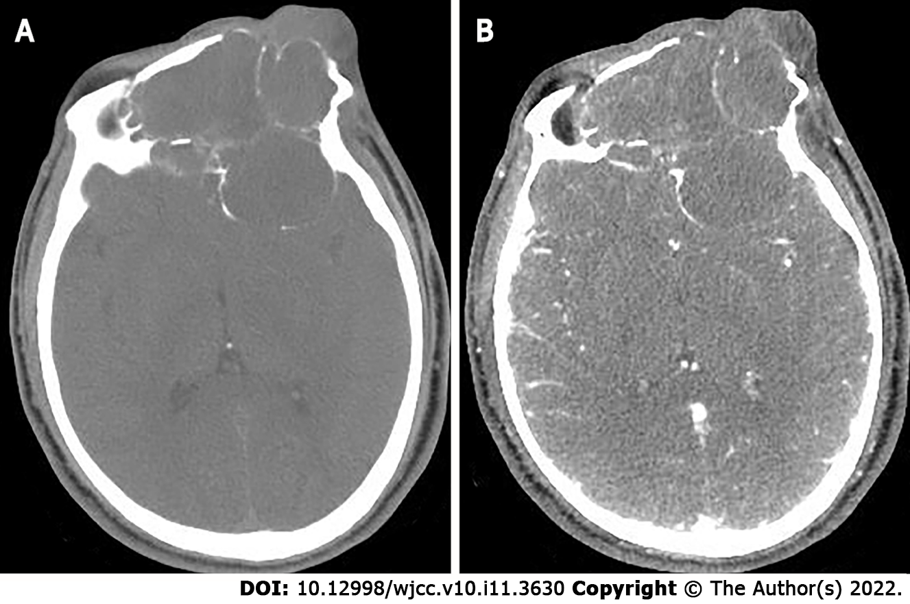
Figure 3 The second preoperative computed tomography findings.
A: Computed tomography (CT) scan shows large lamellar soft tissue density masses in the left orbit, bilateral frontal areas, bilateral septal sinuses, pterygoid sinuses, left maxillary sinus, and anterior skull base, with CT attenuation of approximately 22 Hounsfield units (HU), peripheral multiple osteolytic destruction of bone; B: Arterial phase shows moderate enhancement of the lesion, with CT attenuation of approximately 58 HU.
- Citation: Huang WP, Li LM, Gao JB. Pleomorphic adenoma of the left lacrimal gland recurred and transformed into myoepithelial carcinoma after multiple operations: A case report. World J Clin Cases 2022; 10(11): 3630-3638
- URL: https://www.wjgnet.com/2307-8960/full/v10/i11/3630.htm
- DOI: https://dx.doi.org/10.12998/wjcc.v10.i11.3630


