Copyright
©The Author(s) 2022.
World J Clin Cases. Apr 16, 2022; 10(11): 3630-3638
Published online Apr 16, 2022. doi: 10.12998/wjcc.v10.i11.3630
Published online Apr 16, 2022. doi: 10.12998/wjcc.v10.i11.3630
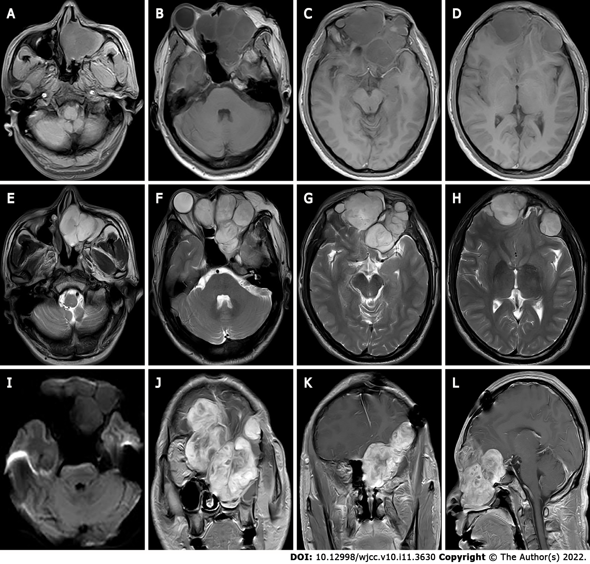
Figure 1 The first preoperative Magnetic resonance imaging findings.
A-I: Magnetic resonance imaging shows multiple masses with T1 weighted imaging (WI) inhomogeneous low signals (A-D) and T2WI inhomogeneous high signals (E-H) in the bilateral frontal, bilateral septal sinuses, bilateral frontal sinuses, left orbit, left nasal cavity, left pterygoid sinus and left maxillary sinus, the lesion showed equal or slightly lower signals on diffusion WI with poorly defined borders (I); J-L: Bilateral frontal lobe and right orbital compression resulted in a localized left shift of the frontal midline, and right shift of the nasal cavity. After enhancement, the lesion showed significant heterogeneous progressive enhancement.
- Citation: Huang WP, Li LM, Gao JB. Pleomorphic adenoma of the left lacrimal gland recurred and transformed into myoepithelial carcinoma after multiple operations: A case report. World J Clin Cases 2022; 10(11): 3630-3638
- URL: https://www.wjgnet.com/2307-8960/full/v10/i11/3630.htm
- DOI: https://dx.doi.org/10.12998/wjcc.v10.i11.3630


