Copyright
©The Author(s) 2022.
World J Clin Cases. Apr 16, 2022; 10(11): 3561-3572
Published online Apr 16, 2022. doi: 10.12998/wjcc.v10.i11.3561
Published online Apr 16, 2022. doi: 10.12998/wjcc.v10.i11.3561
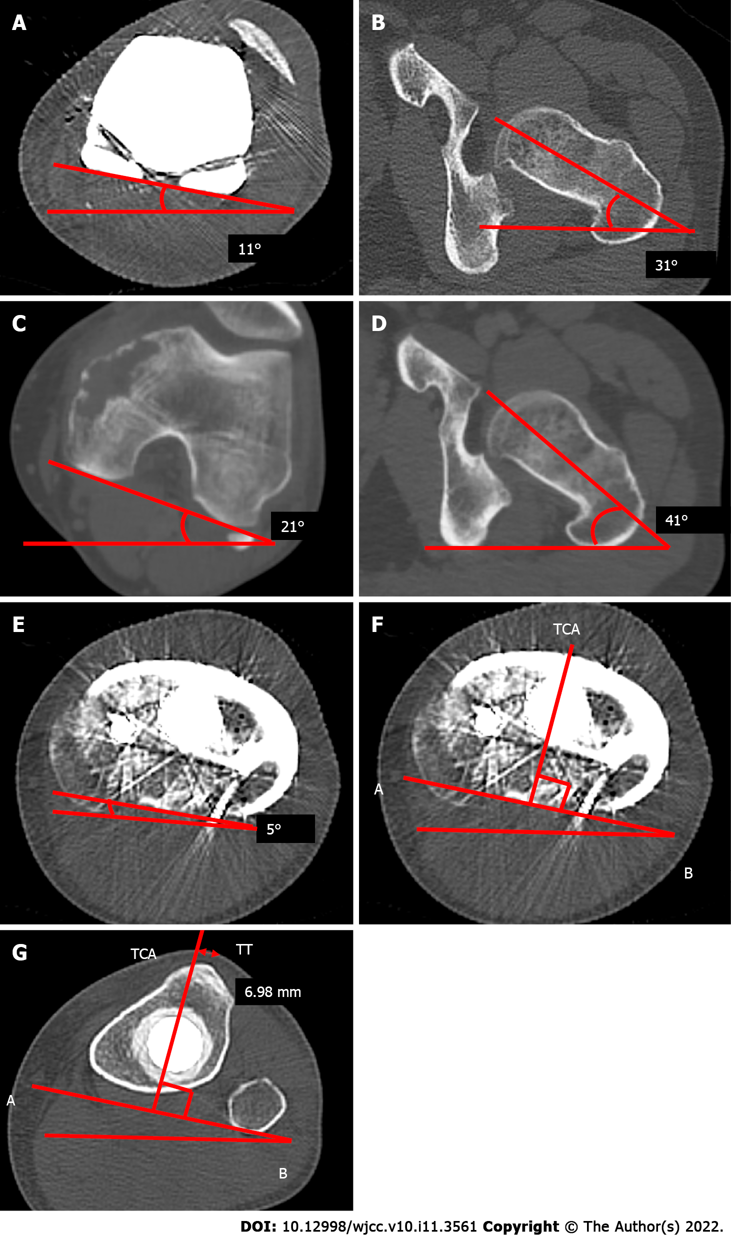
Figure 3 The computed tomography of a femoral axial and tibial axial view after or before the primary operation.
A-D: The computed tomography (CT) imaging of a femoral axial after (A and B) and before (C and D) the primary operation, which shows that the postoperative angle between the femoral component and the femoral neck axis (A and B) is equal to the preoperative angle between the femoral posterior condylar axis and femoral neck axis (C and D); E-G: A CT scan shows the axial rotation of the tibial component in relation to the posterior margins of the tibial plateau and the tibial bearing after the primary operation. The line AB is drawn along the posterior margin of the tibial tray. The tibial component axis (TCA) is perpendicular to line AB (E and F); the perpendicular distance from the TCA to the tip of the tibial tuberosity is 6.98 mm (G). TCA: Tibial component axis; TT: Tibial tuberosity.
- Citation: Kubota Y, Tanaka K, Hirakawa M, Iwasaki T, Kawano M, Itonaga I, Tsumura H. Patellar dislocation following distal femoral replacement after extra-articular knee resection for bone sarcoma: A case report. World J Clin Cases 2022; 10(11): 3561-3572
- URL: https://www.wjgnet.com/2307-8960/full/v10/i11/3561.htm
- DOI: https://dx.doi.org/10.12998/wjcc.v10.i11.3561


