Copyright
©The Author(s) 2022.
World J Clin Cases. Apr 16, 2022; 10(11): 3547-3552
Published online Apr 16, 2022. doi: 10.12998/wjcc.v10.i11.3547
Published online Apr 16, 2022. doi: 10.12998/wjcc.v10.i11.3547
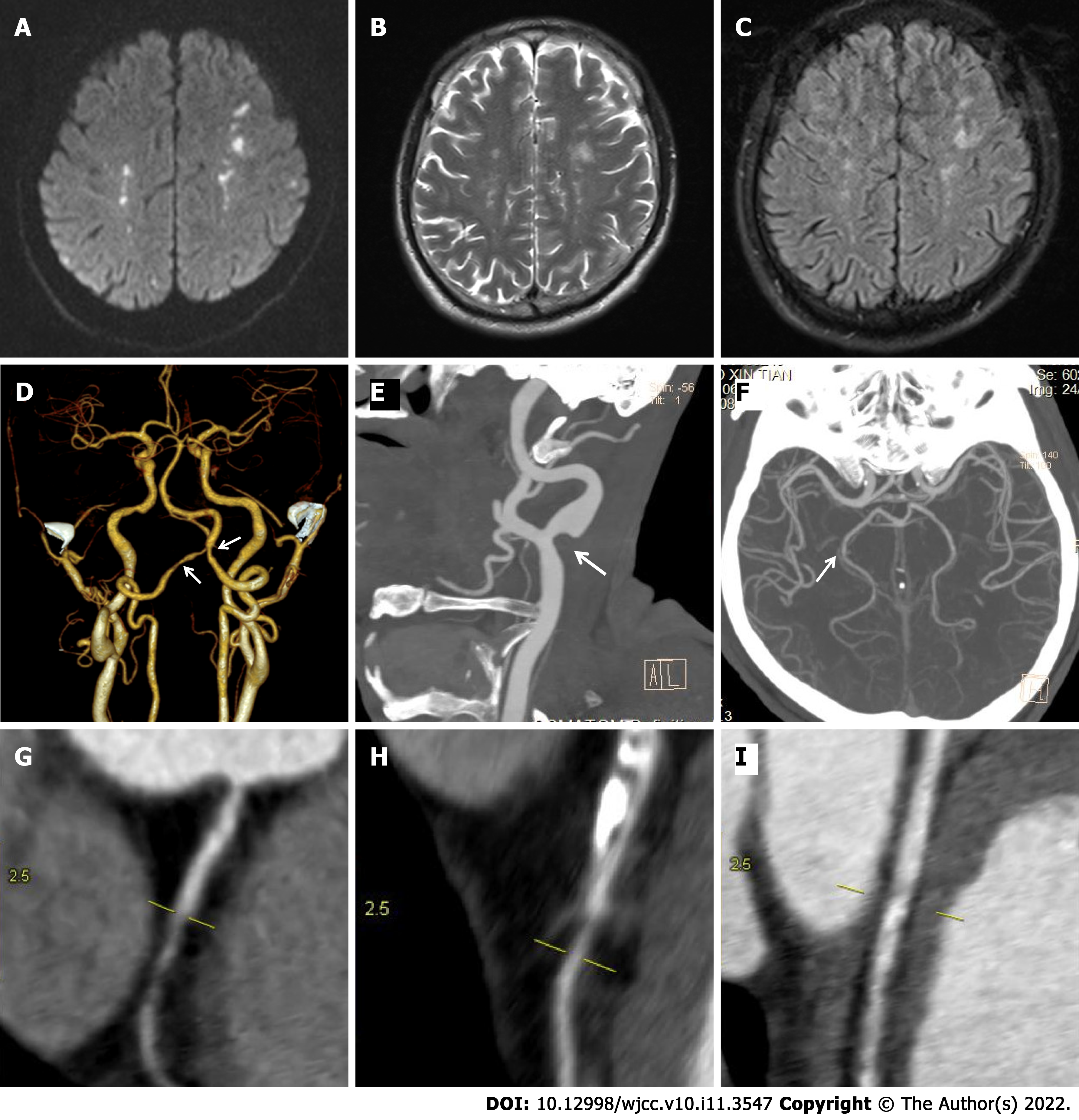
Figure 1 Brain magnetic resonance imaging and computed tomography angiography at admission.
A: Diffusion-weighted magnetic resonance imaging (MRI); B: T2-weighted MRI; C: Fluid-attenuated inversion recovery revealed multiple acute infarcts in the bilateral border zones; D-I: Computed tomography angiogram (CTA) of the head and neck revealed mild stenosis in the (D) bilateral vertebral arteries, (E) left common carotid artery bifurcation, and (F) the posterior cerebral artery. Coronary CTA showed mild stenosis in the (G) right coronary artery, (H) left anterior descending artery, and (I) the left circumflex artery.
- Citation: Sun RR, Chen TZ, Meng M. Hypereosinophilic syndrome presenting as acute ischemic stroke, myocardial infarction, and arterial involvement: A case report. World J Clin Cases 2022; 10(11): 3547-3552
- URL: https://www.wjgnet.com/2307-8960/full/v10/i11/3547.htm
- DOI: https://dx.doi.org/10.12998/wjcc.v10.i11.3547


