Copyright
©The Author(s) 2022.
World J Clin Cases. Apr 16, 2022; 10(11): 3518-3526
Published online Apr 16, 2022. doi: 10.12998/wjcc.v10.i11.3518
Published online Apr 16, 2022. doi: 10.12998/wjcc.v10.i11.3518
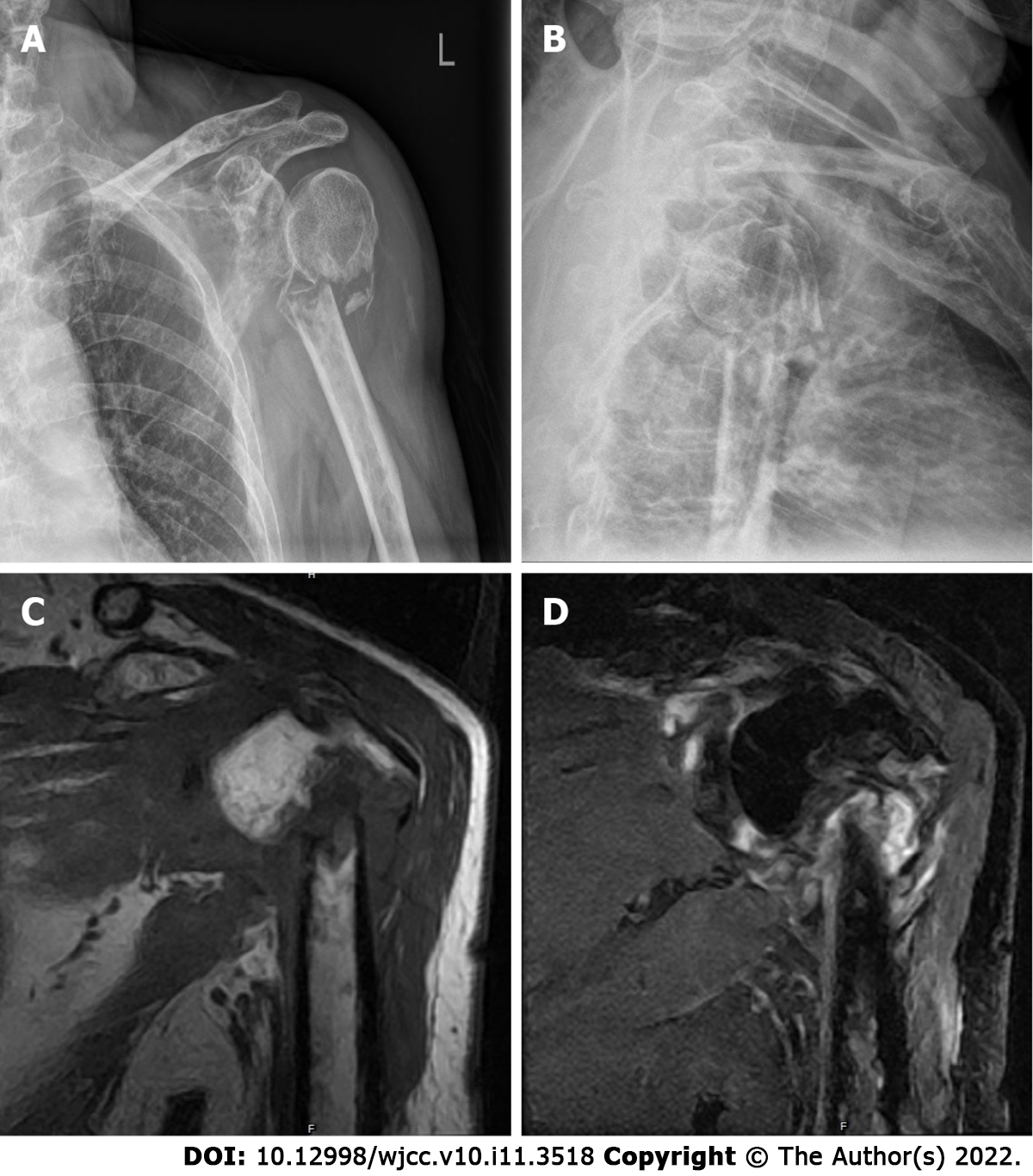
Figure 2 Preoperative X-ray films and magnetic resonance imaging.
A, B: X-ray films (A, B) of the left shoulder revealed only a comminuted fracture and degenerative changes in the upper left humerus; C, D: Multiple soft tissue masses around the fracture presented as hypointensity and isointensity on T1 weighted (T1-W) images (C) and hybrid hyperintensity on T2-W images (D).
- Citation: Xu GQ, Wang G, Bai XD, Wang XJ. Intramedullary nailing for pathological fractures of the proximal humerus caused by multiple myeloma: A case report and review of literature. World J Clin Cases 2022; 10(11): 3518-3526
- URL: https://www.wjgnet.com/2307-8960/full/v10/i11/3518.htm
- DOI: https://dx.doi.org/10.12998/wjcc.v10.i11.3518


