Copyright
©The Author(s) 2022.
World J Clin Cases. Apr 16, 2022; 10(11): 3485-3489
Published online Apr 16, 2022. doi: 10.12998/wjcc.v10.i11.3485
Published online Apr 16, 2022. doi: 10.12998/wjcc.v10.i11.3485
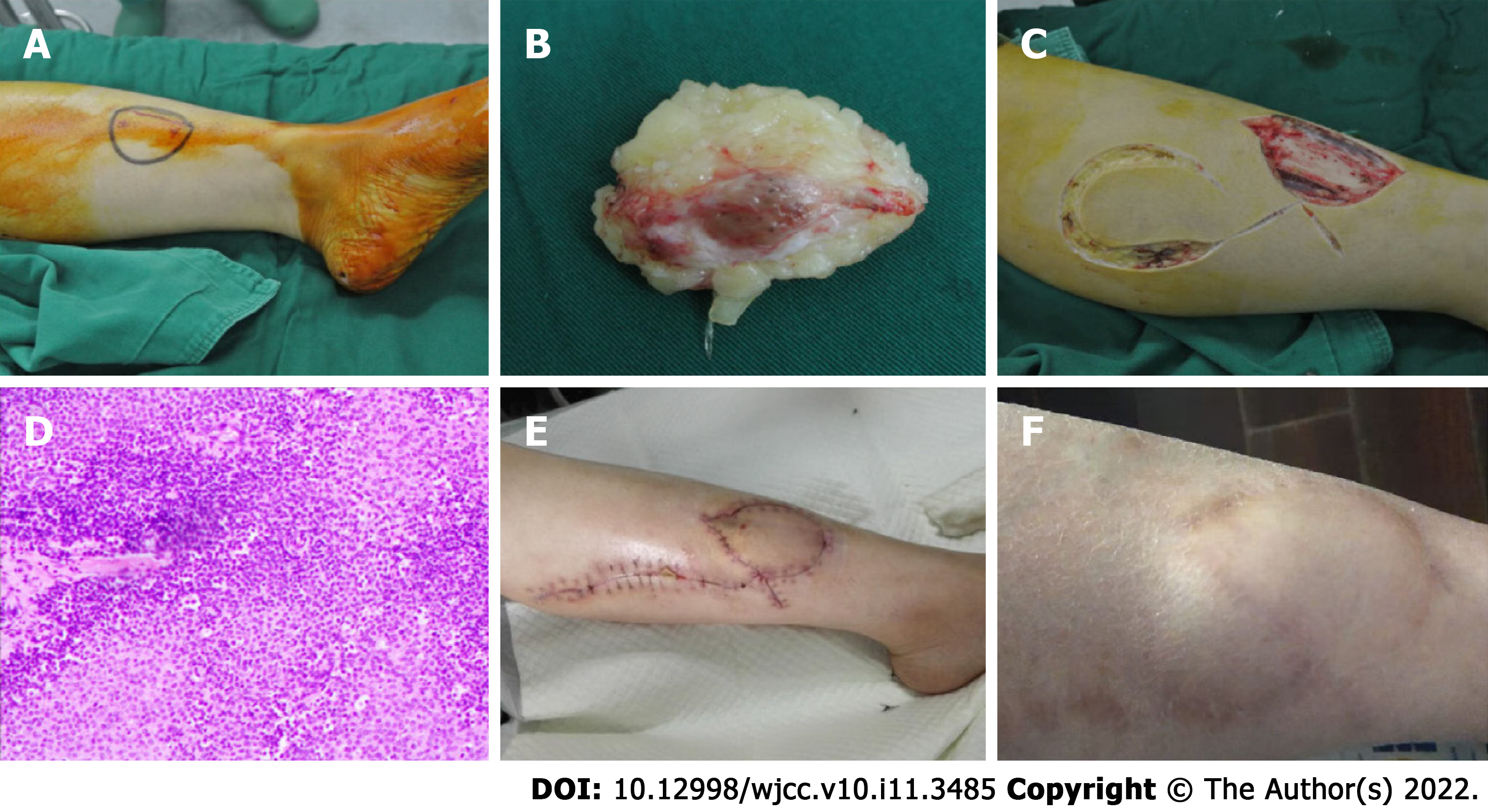
Figure 1 The imaging of case.
A: Surgical field: The appearance of the medial left lower leg; B: The appearance of excised tissue in operation. Ellipsoid red soft tissue be covered with adipose tissue, about 50 mm × 30 mm ×10 mm in size; C: Designing and harvesting the lateral gastrocnemius nutrition flap of the left lower leg to cover the tumor's resection site during the surgery; D: Pathologic examination shows a glomus tumor with chronic inflammatory cell infiltrates; E: The appearance of the anteromedial incision of the left lower leg two weeks after the surgery. The incision suture had been removed. The flap survived well, and the incision recovered well with no infection; F: The anteromedial appearance of the left lower leg four years after the surgery. The pain did not relapse.
- Citation: Wang HY, Duan P, Chen H, Pan ZY. Unusual glomus tumor of the lower leg: A case report. World J Clin Cases 2022; 10(11): 3485-3489
- URL: https://www.wjgnet.com/2307-8960/full/v10/i11/3485.htm
- DOI: https://dx.doi.org/10.12998/wjcc.v10.i11.3485


