Copyright
©The Author(s) 2022.
World J Clin Cases. Apr 16, 2022; 10(11): 3472-3477
Published online Apr 16, 2022. doi: 10.12998/wjcc.v10.i11.3472
Published online Apr 16, 2022. doi: 10.12998/wjcc.v10.i11.3472
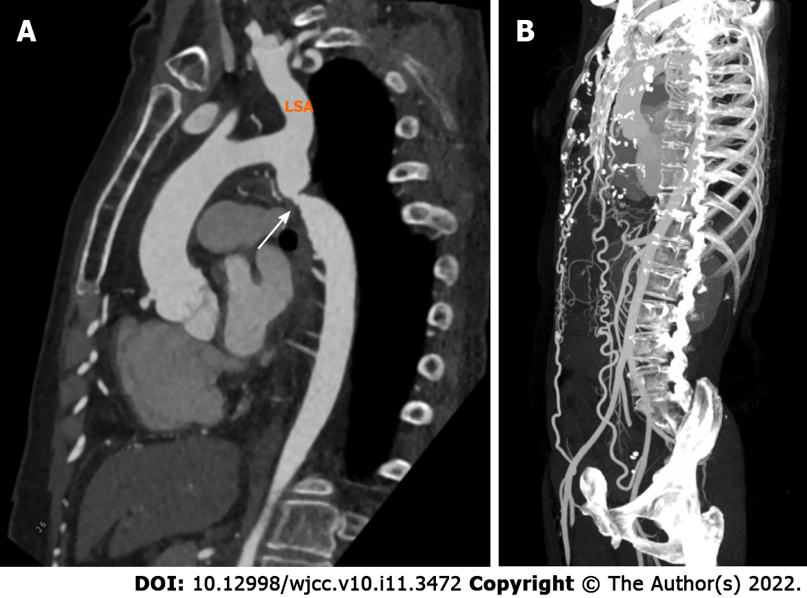
Figure 2 Severe coarctation in a 61-year-old woman.
A: Reformatted oblique sagittal image of the aorta shows coarctation of the aorta just distal to the subclavian artery origin (white arrow) and slight enlargement of the proximal descending aorta. The ascending aorta maintains normal curvature and the narrowing is a few centimeters away from the subclavian artery; B: Maximum intensity projection shows collateral flow which included two full-fledged collateral networks ensuring blood circulation to the distal aorta. LSA: Left subclavian artery.
- Citation: Ren SX, Zhang Q, Li PP, Wang XD. Difference and similarity between type A interrupted aortic arch and aortic coarctation in adults: Two case reports. World J Clin Cases 2022; 10(11): 3472-3477
- URL: https://www.wjgnet.com/2307-8960/full/v10/i11/3472.htm
- DOI: https://dx.doi.org/10.12998/wjcc.v10.i11.3472


