Copyright
©The Author(s) 2022.
World J Clin Cases. Apr 16, 2022; 10(11): 3369-3378
Published online Apr 16, 2022. doi: 10.12998/wjcc.v10.i11.3369
Published online Apr 16, 2022. doi: 10.12998/wjcc.v10.i11.3369
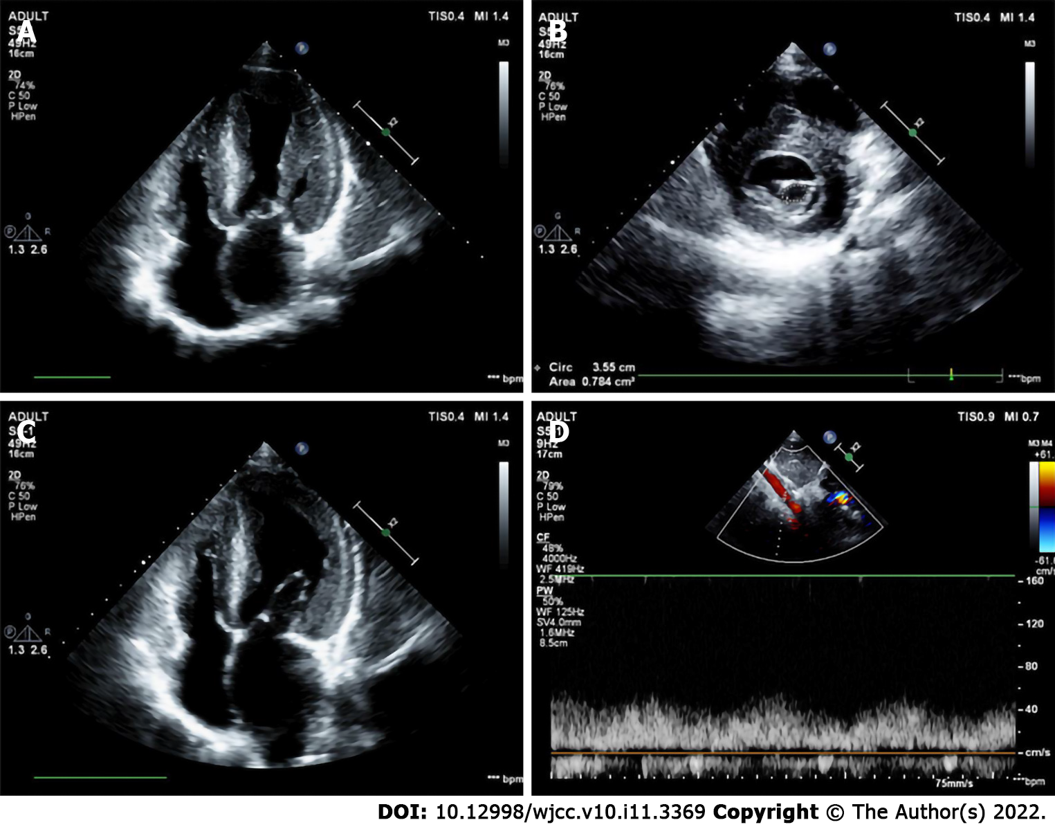
Figure 3 A case in which the Annulo-Leaflet mitral ring was missed by echocardiography at the outpatient.
Intraoperative exploration revealed that the supramitral ring was adhered to the anterior and posterior leaflets, close to the anterior and posterior leaflets of the mitral valve. A: The parasternal mitral short-axis view showed a significant reduction in the area of the mitral valve orifice, which was only 0.78 cm2; B: Two-dimensional ultrasonography at the apical four-chamber view showed thickened mitral valve with restricted opening; C: There are both annulo-leaflet mitral ring and parachute mitral valve. Preoperative echocardiography after admission showed a tiny septum on the anterior leaflet of the mitral valve; D: Spectral Doppler of the abdominal aorta showed low velocity and low resistance, suggesting coarctation of the aorta.
- Citation: Li YD, Meng H, Pang KJ, Li MZ, Xu N, Wang H, Li SJ, Yan J. Echocardiography in the diagnosis of Shone’s complex and analysis of the causes for missed diagnosis and misdiagnosis. World J Clin Cases 2022; 10(11): 3369-3378
- URL: https://www.wjgnet.com/2307-8960/full/v10/i11/3369.htm
- DOI: https://dx.doi.org/10.12998/wjcc.v10.i11.3369


