Copyright
©The Author(s) 2022.
World J Clin Cases. Apr 16, 2022; 10(11): 3369-3378
Published online Apr 16, 2022. doi: 10.12998/wjcc.v10.i11.3369
Published online Apr 16, 2022. doi: 10.12998/wjcc.v10.i11.3369
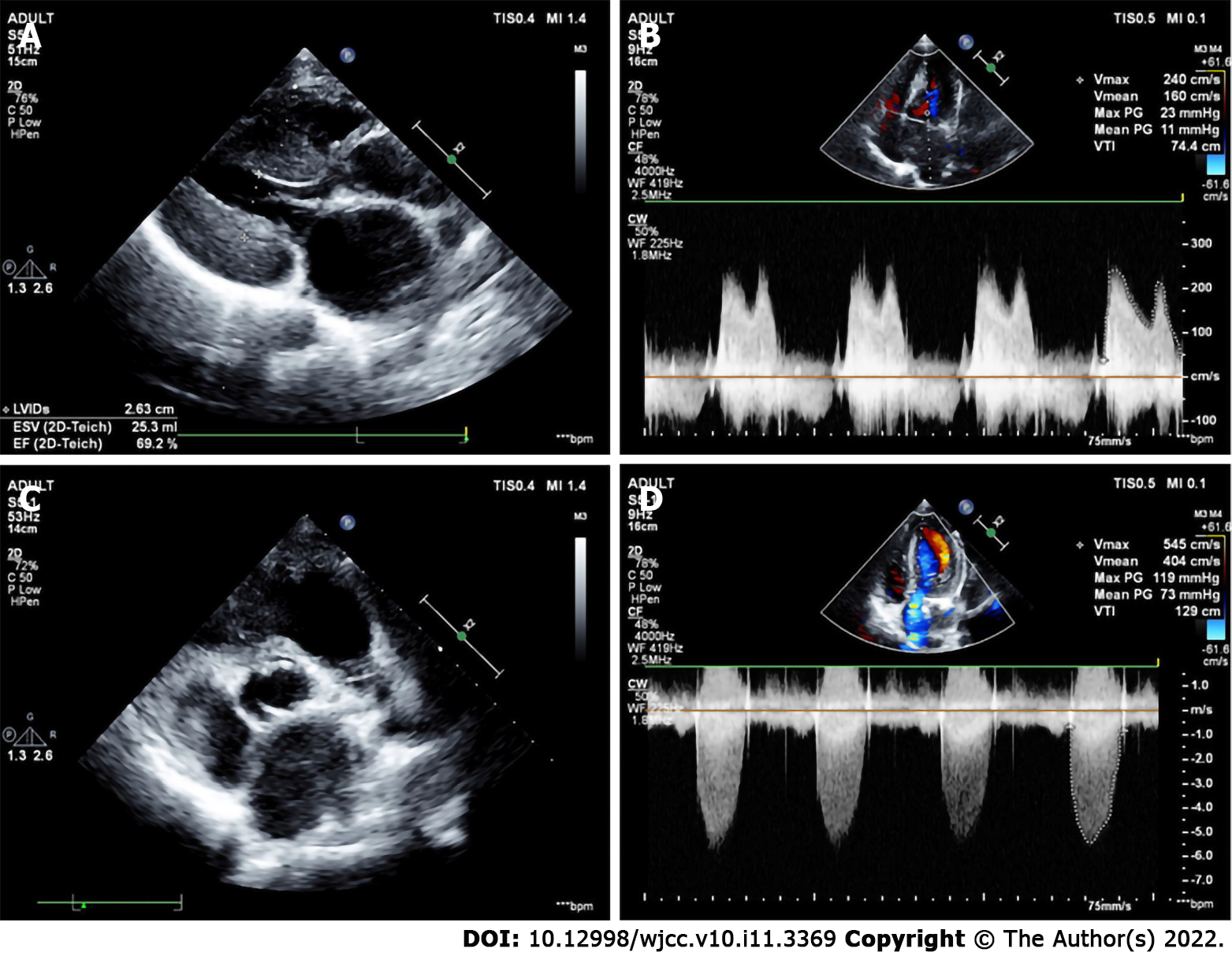
Figure 2 Image of a patient with incomplete Shone’s complex.
Echocardiography missed the ALMR. Intraoperative exploration revealed that the mitral valve adhered to the anterior leaflet, close to the anterior leaflet of the mitral valve. A: The parasternal left ventricular long-axis view showed a significant thickening of the left ventricular wall; B: The forward flow velocity at the mitral valve was increased significantly, at 2.4 m/s, and the mean transvalvular pressure gradient was 11 mmHg; C: The aortic valve was bicuspid and arranged on the left and right; D: The flow velocity of the aortic valve was increased significantly, at 5.45 m/s, and the mean transvalvular pressure gradient was 73 mmHg.
- Citation: Li YD, Meng H, Pang KJ, Li MZ, Xu N, Wang H, Li SJ, Yan J. Echocardiography in the diagnosis of Shone’s complex and analysis of the causes for missed diagnosis and misdiagnosis. World J Clin Cases 2022; 10(11): 3369-3378
- URL: https://www.wjgnet.com/2307-8960/full/v10/i11/3369.htm
- DOI: https://dx.doi.org/10.12998/wjcc.v10.i11.3369


