Copyright
©The Author(s) 2022.
World J Clin Cases. Apr 6, 2022; 10(10): 3313-3320
Published online Apr 6, 2022. doi: 10.12998/wjcc.v10.i10.3313
Published online Apr 6, 2022. doi: 10.12998/wjcc.v10.i10.3313
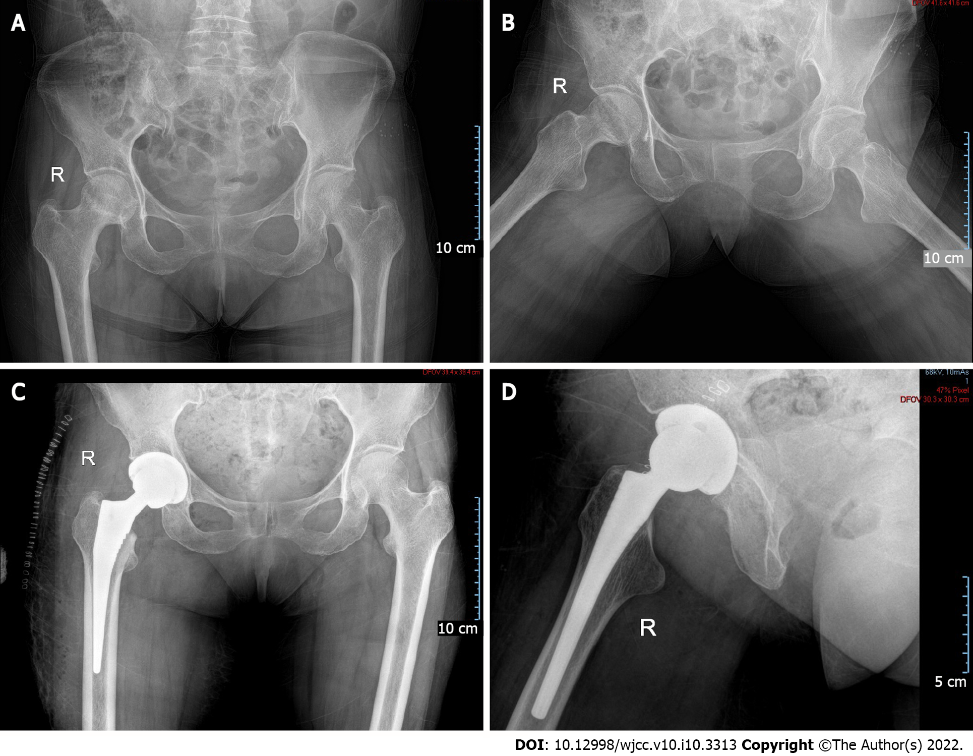
Figure 1 Hip imaging findings.
A and B: Preoperative anterior and lateral X-ray films of both hips, showing the uneven density of the femoral head; C and D: Postoperative anterior and lateral X-ray films of the right hip, showing good prosthesis positioning.
- Citation: Hong M, Zhang ZY, Sun XW, Wang WG, Zhang QD, Guo WS. Pneumocystis jiroveci pneumonia after total hip arthroplasty in a dermatomyositis patient: A case report . World J Clin Cases 2022; 10(10): 3313-3320
- URL: https://www.wjgnet.com/2307-8960/full/v10/i10/3313.htm
- DOI: https://dx.doi.org/10.12998/wjcc.v10.i10.3313


