Copyright
©The Author(s) 2022.
World J Clin Cases. Apr 6, 2022; 10(10): 3297-3305
Published online Apr 6, 2022. doi: 10.12998/wjcc.v10.i10.3297
Published online Apr 6, 2022. doi: 10.12998/wjcc.v10.i10.3297
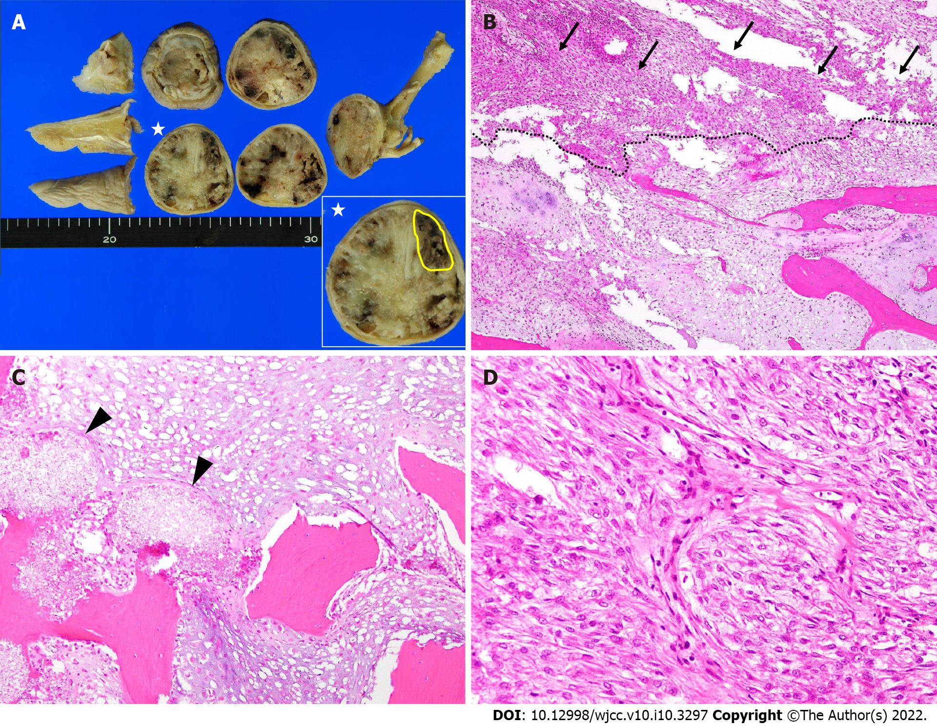
Figure 4 Histological findings of the amputated specimen.
A: Macroscopically, the tumor has destroyed the bone cortex and is extended to the soft tissue below the skin. The tumor is a grayish-white hyaline cartilage component filling the medullary cavity with hemorrhage and necrosis. In the largest cross-section of the tumor (star), the high-grade dedifferentiated component (yellow) accounts for 10.2% of the tumor; B: Magnification × 4. There is an abrupt transition between the low-grade chondrosarcoma (lower side) and high-grade dedifferentiated sarcoma components (upper side; arrow); C: Magnification × 10. The cartilaginous portion of the tumor shows a grade 1 or grade 2 chondrosarcoma with bone destruction. Arrowheads show the hydroxyapatite artificial bone graft that was used 11 years ago; D: Magnification × 20. The high-grade dedifferentiated component shows mitoses and atypia. The lesion is composed of spindle cells arranged in a fascicular growth pattern, resembling a high-grade undifferentiated pleomorphic sarcoma.
- Citation: Yonezawa H, Yamamoto N, Hayashi K, Takeuchi A, Miwa S, Igarashi K, Morinaga S, Asano Y, Saito S, Tome Y, Ikeda H, Nojima T, Tsuchiya H. Dedifferentiated chondrosarcoma of the middle finger arising from a solitary enchondroma: A case report. World J Clin Cases 2022; 10(10): 3297-3305
- URL: https://www.wjgnet.com/2307-8960/full/v10/i10/3297.htm
- DOI: https://dx.doi.org/10.12998/wjcc.v10.i10.3297


