Copyright
©The Author(s) 2022.
World J Clin Cases. Apr 6, 2022; 10(10): 3297-3305
Published online Apr 6, 2022. doi: 10.12998/wjcc.v10.i10.3297
Published online Apr 6, 2022. doi: 10.12998/wjcc.v10.i10.3297
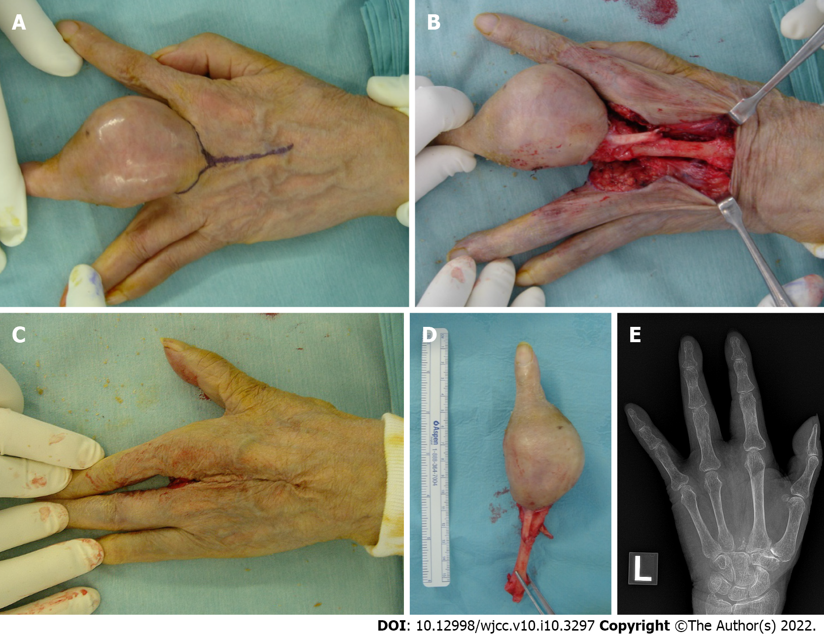
Figure 3 Intraoperative photographs and postoperative X-ray of the second surgery at 87 years of age.
A: A racket-shaped incision was planned; B: The third metacarpal bone is exposed and disarticulated at the carpometacarpal joint; C: The postoperative photograph of the dorsal aspect of the left hand demonstrates the gap closure; D: The amputated specimen; E: The postoperative X-ray of the left hand shows a good cosmetic four-finger hand.
- Citation: Yonezawa H, Yamamoto N, Hayashi K, Takeuchi A, Miwa S, Igarashi K, Morinaga S, Asano Y, Saito S, Tome Y, Ikeda H, Nojima T, Tsuchiya H. Dedifferentiated chondrosarcoma of the middle finger arising from a solitary enchondroma: A case report. World J Clin Cases 2022; 10(10): 3297-3305
- URL: https://www.wjgnet.com/2307-8960/full/v10/i10/3297.htm
- DOI: https://dx.doi.org/10.12998/wjcc.v10.i10.3297


