Copyright
©The Author(s) 2022.
World J Clin Cases. Apr 6, 2022; 10(10): 3297-3305
Published online Apr 6, 2022. doi: 10.12998/wjcc.v10.i10.3297
Published online Apr 6, 2022. doi: 10.12998/wjcc.v10.i10.3297
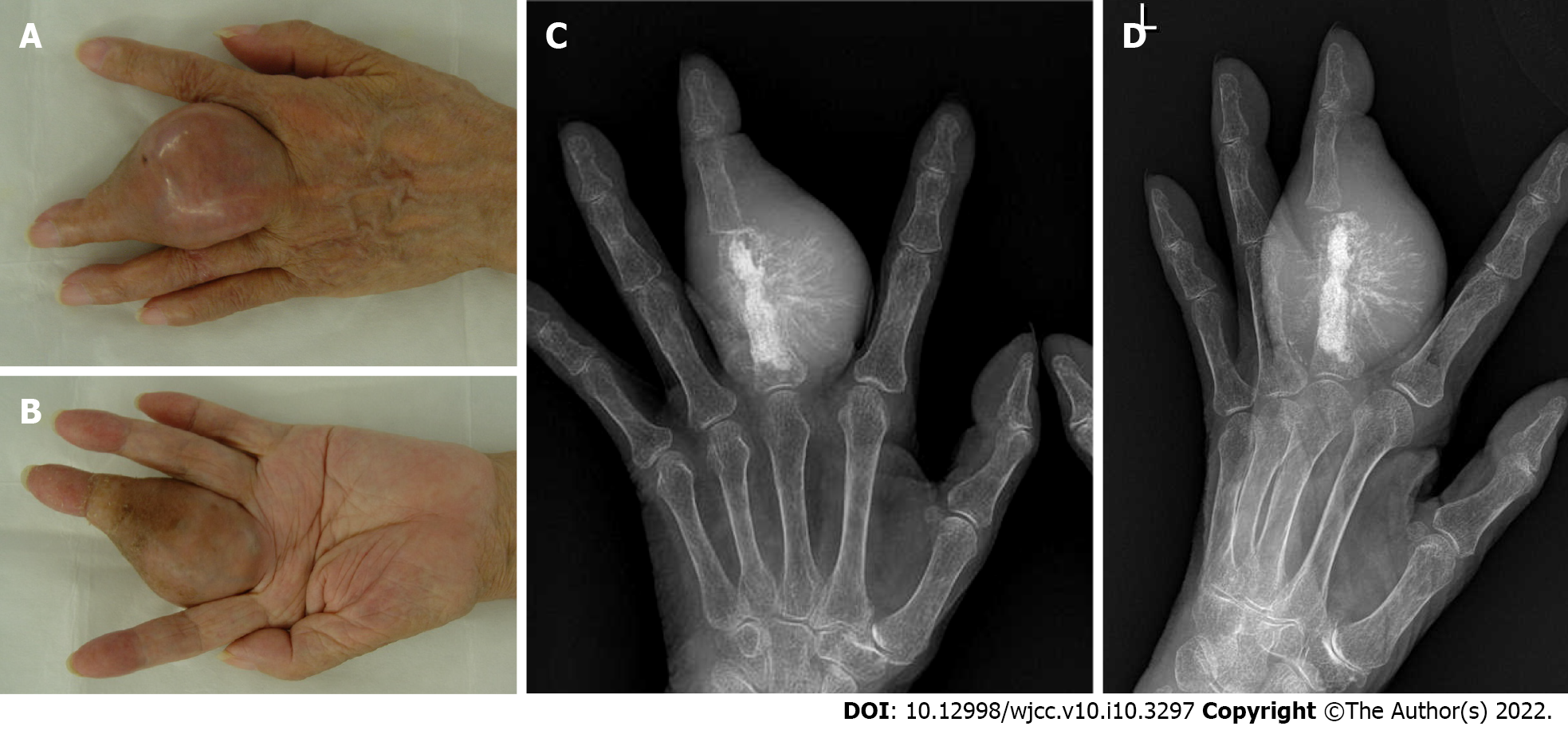
Figure 2 Clinical photographs and X-ray of the finger 11 years after the primary surgery.
A and B: Clinical photographs of the hand at 87 years of age. The left middle finger appears swollen and tense. The scar of the primary surgery is observed in the dorsal surface of the middle finger; A: Dorsal view; B: Palmar view; C and D: The X-ray shows residual artificial bone graft material and an expansive osteolytic lesion in the proximal phalanx of the middle finger; C: Anteroposterior view; D: Oblique view.
- Citation: Yonezawa H, Yamamoto N, Hayashi K, Takeuchi A, Miwa S, Igarashi K, Morinaga S, Asano Y, Saito S, Tome Y, Ikeda H, Nojima T, Tsuchiya H. Dedifferentiated chondrosarcoma of the middle finger arising from a solitary enchondroma: A case report. World J Clin Cases 2022; 10(10): 3297-3305
- URL: https://www.wjgnet.com/2307-8960/full/v10/i10/3297.htm
- DOI: https://dx.doi.org/10.12998/wjcc.v10.i10.3297


