Copyright
©The Author(s) 2022.
World J Clin Cases. Apr 6, 2022; 10(10): 3297-3305
Published online Apr 6, 2022. doi: 10.12998/wjcc.v10.i10.3297
Published online Apr 6, 2022. doi: 10.12998/wjcc.v10.i10.3297
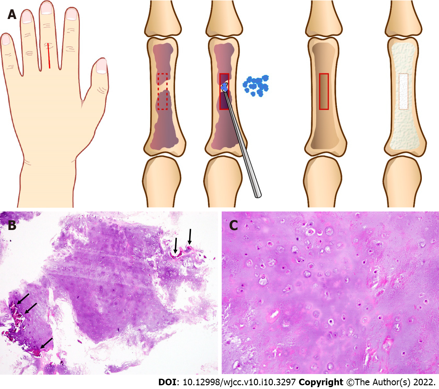
Figure 1 Schema and histological findings of the primary surgery at 76 years of age.
A: Schema of the primary surgery referencing the surgical record and figures. The tumor is located in the third proximal phalanx. After fenestration of the bone cortex, curettage and granular artificial bone grafting were performed under axillary nerve block. B: Magnification × 4. The tumor appears as a lobular, relatively cell-poor hyaline cartilage surrounded by an eosinophilic zone of reactive bone formation (encasement pattern; arrow). C: Magnification × 20. Scattered chondrocytes are located in sharp-edged lacunar spaces, with abundant hyaline cartilage matrix. Nuclei are small, round, and hyperchromatic, although larger, vesicular nuclei can also be present. No nuclear pleomorphism or enlargement is observed.
- Citation: Yonezawa H, Yamamoto N, Hayashi K, Takeuchi A, Miwa S, Igarashi K, Morinaga S, Asano Y, Saito S, Tome Y, Ikeda H, Nojima T, Tsuchiya H. Dedifferentiated chondrosarcoma of the middle finger arising from a solitary enchondroma: A case report. World J Clin Cases 2022; 10(10): 3297-3305
- URL: https://www.wjgnet.com/2307-8960/full/v10/i10/3297.htm
- DOI: https://dx.doi.org/10.12998/wjcc.v10.i10.3297


