Copyright
©The Author(s) 2022.
World J Clin Cases. Apr 6, 2022; 10(10): 3291-3296
Published online Apr 6, 2022. doi: 10.12998/wjcc.v10.i10.3291
Published online Apr 6, 2022. doi: 10.12998/wjcc.v10.i10.3291
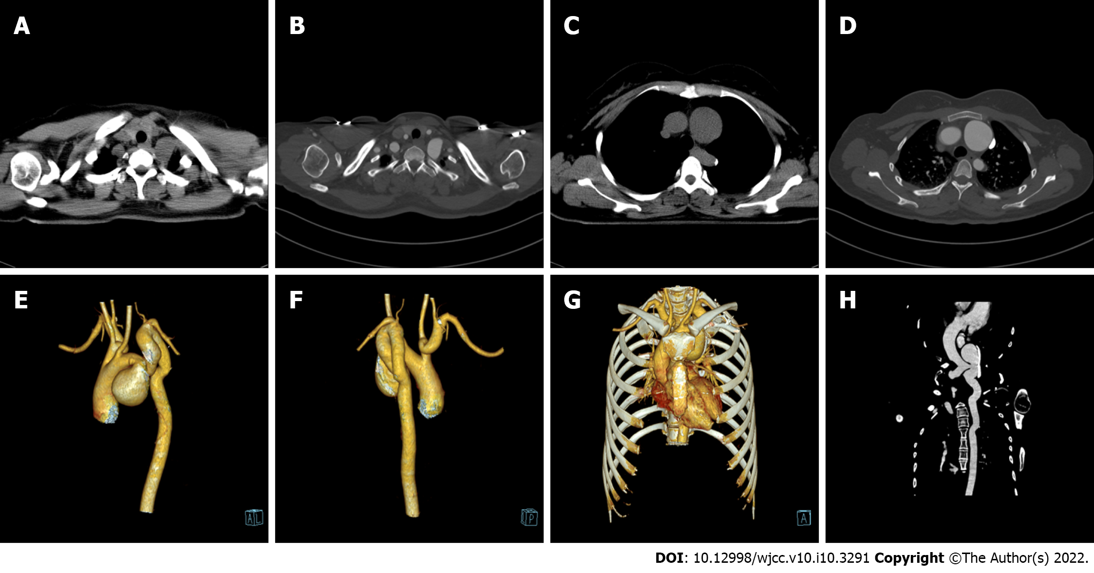
Figure 1 Cervical aortic arch in a 29-year-old female patient.
A: Non-contrast-enhanced chest computed tomography (CT) images (axial view) showing that the top of the aortic arch (AA) was at the mid-superior level of the first thoracic vertebra; B: Contrast-enhanced CT images (axial view) showing the same sign as A; C: Non-contrast-enhanced chest CT images (axial view) showing that the largest plane of the aortic aneurysm was roughly the upper plane of the fifth thoracic vertebrae; D: Contrast-enhanced CT images (axial view) showing the same sign as C; E: Volume reconstruction (VR) (left anterior oblique view) showing that the brachiocephalic trunk, left common carotid artery, and left subclavian artery were sequential branches from the proximal end to the distal end of the AA, that the AA showed tumor-like local expansion, and that the AA distal to the aneurysm and the upper descending aorta (DA) were long and tortuous; F: VR (left posterior oblique view) showing that the right vertebral artery (RVA) originated from the right subclavian artery (RSCA), the left vertebral artery (LVA) originated from the top of the AA, the LVA was thinner than the RVA, and the proximal segment of the RSCA was dilated and tortuous; G: VR (anteroposterior view) showing that the top of the AA was located above the clavicle; H: Curved planar reconstruction showing that the AA straddled the left side of the spine and that the AA distal to the aneurysm and the upper segment of the DA were long and tortuous and were located to the left of the spine.
- Citation: Wu YK, Mao Q, Zhou MT, Liu N, Yu X, Peng JC, Tao YY, Gong XQ, Yang L, Zhang XM. Cervical aortic arch with aneurysm formation and an anomalous right subclavian artery and left vertebral artery: A case report. World J Clin Cases 2022; 10(10): 3291-3296
- URL: https://www.wjgnet.com/2307-8960/full/v10/i10/3291.htm
- DOI: https://dx.doi.org/10.12998/wjcc.v10.i10.3291


