Copyright
©The Author(s) 2022.
World J Clin Cases. Apr 6, 2022; 10(10): 3251-3260
Published online Apr 6, 2022. doi: 10.12998/wjcc.v10.i10.3251
Published online Apr 6, 2022. doi: 10.12998/wjcc.v10.i10.3251
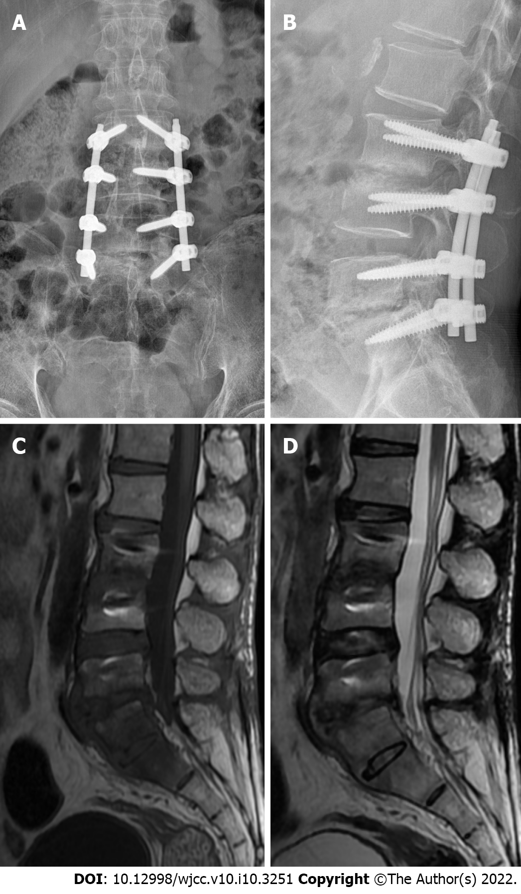
Figure 3 Medical imaging examinations at follow-up.
A and B: X-ray image showing that the position of fixation in the L3–S1 vertebral body was good; C and D: Magnetic resonance imaging showing increased density of the intervertebral spaces of L3/L4 and L5/S1, with a weak T1 signal. No recurrence was noted.
- Citation: Shi XW, Li ST, Lou JP, Xu B, Wang J, Wang X, Liu H, Li SK, Zhen P, Zhang T. Scedosporium apiospermum infection of the lumbar vertebrae: A case report. World J Clin Cases 2022; 10(10): 3251-3260
- URL: https://www.wjgnet.com/2307-8960/full/v10/i10/3251.htm
- DOI: https://dx.doi.org/10.12998/wjcc.v10.i10.3251


