Copyright
©The Author(s) 2022.
World J Clin Cases. Apr 6, 2022; 10(10): 3241-3250
Published online Apr 6, 2022. doi: 10.12998/wjcc.v10.i10.3241
Published online Apr 6, 2022. doi: 10.12998/wjcc.v10.i10.3241
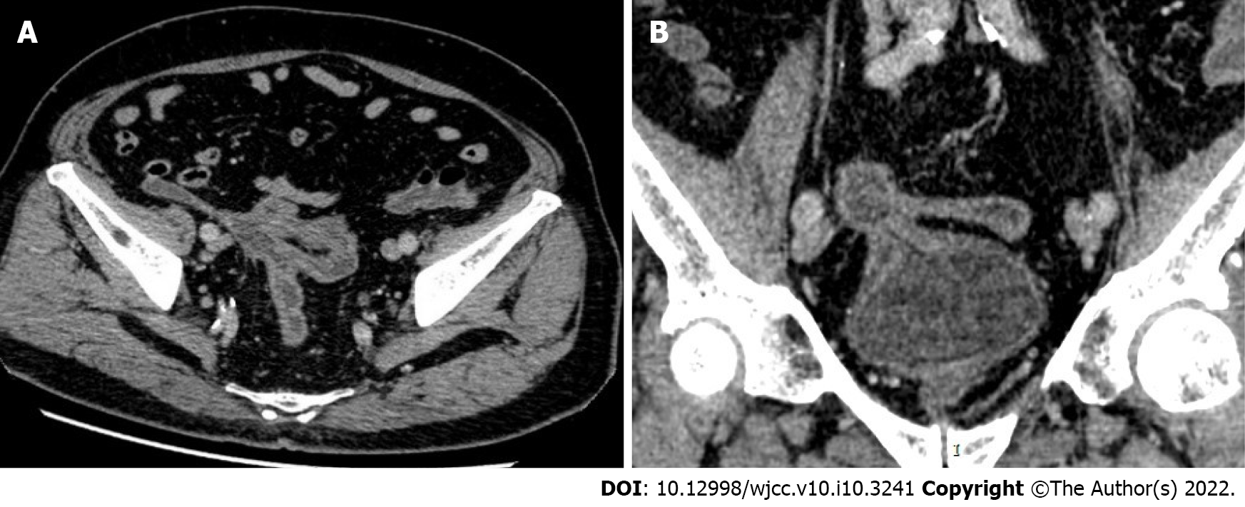
Figure 2 Contrast-enhanced computed tomography of the abdominal pelvis: the appendix was not clearly visualized.
A: The distal sigmoid colon-cecum, right posterior to the top wall of the bladder - sigmoid colon, and middle sigmoid colon-cecal wall were adhered, and fistulas could be seen among these structures. The correlative intestinal wall was thickened, and a contrast-enhanced computed tomography (CT) scan showed enhancement; B: A gas density shadow could be seen in the bladder. The correlative bladder wall was thickened, and a contrast-enhanced CT scan showed enhancement.
- Citation: Yan H, Wu YC, Wang X, Liu YC, Zuo S, Wang PY. Appendico-vesicocolonic fistula: A case report and review of literature. World J Clin Cases 2022; 10(10): 3241-3250
- URL: https://www.wjgnet.com/2307-8960/full/v10/i10/3241.htm
- DOI: https://dx.doi.org/10.12998/wjcc.v10.i10.3241


