Copyright
©The Author(s) 2022.
World J Clin Cases. Apr 6, 2022; 10(10): 3206-3212
Published online Apr 6, 2022. doi: 10.12998/wjcc.v10.i10.3206
Published online Apr 6, 2022. doi: 10.12998/wjcc.v10.i10.3206
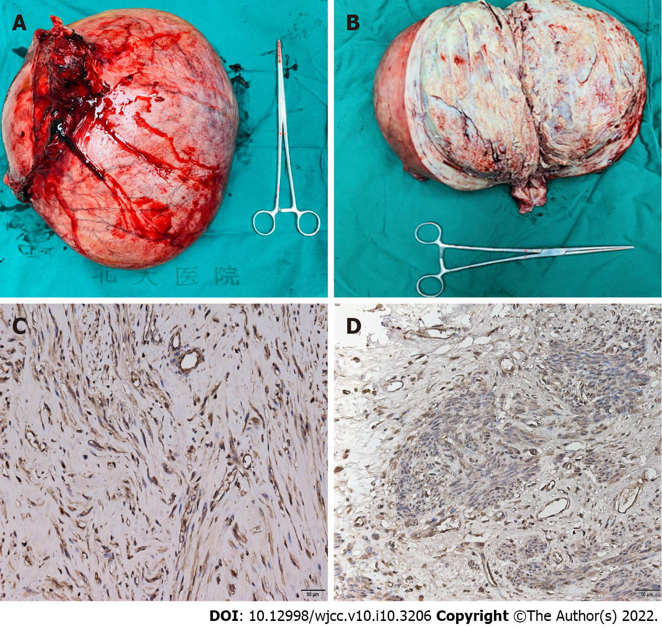
Figure 2 Gross and microscopic analysis of the uterus.
A: The uterus weighed 6500 g, with the largest diameter of 35 cm; B: The sagittal plane of the uterus showed that the large myoma originated from the posterior wall of the uterus, with the cut surface appearing yellow white with scattered red hemorrhagic areas; C: Under the microscope (200 ×), via immunohistochemical staining, the cytoplasm of the leiomyoma cells of this patient showed strong positivity for erythropoietin; D: Compared with the myoma of this patient, the myoma of a patient without erythrocytosis showed weaker erythropoietin staining in immunohistochemical analysis (200 ×).
- Citation: Shu XY, Chen N, Chen BY, Yang HX, Bi H. Myomatous erythrocytosis syndrome: A case report. World J Clin Cases 2022; 10(10): 3206-3212
- URL: https://www.wjgnet.com/2307-8960/full/v10/i10/3206.htm
- DOI: https://dx.doi.org/10.12998/wjcc.v10.i10.3206


