Copyright
©The Author(s) 2022.
World J Clin Cases. Apr 6, 2022; 10(10): 3194-3199
Published online Apr 6, 2022. doi: 10.12998/wjcc.v10.i10.3194
Published online Apr 6, 2022. doi: 10.12998/wjcc.v10.i10.3194
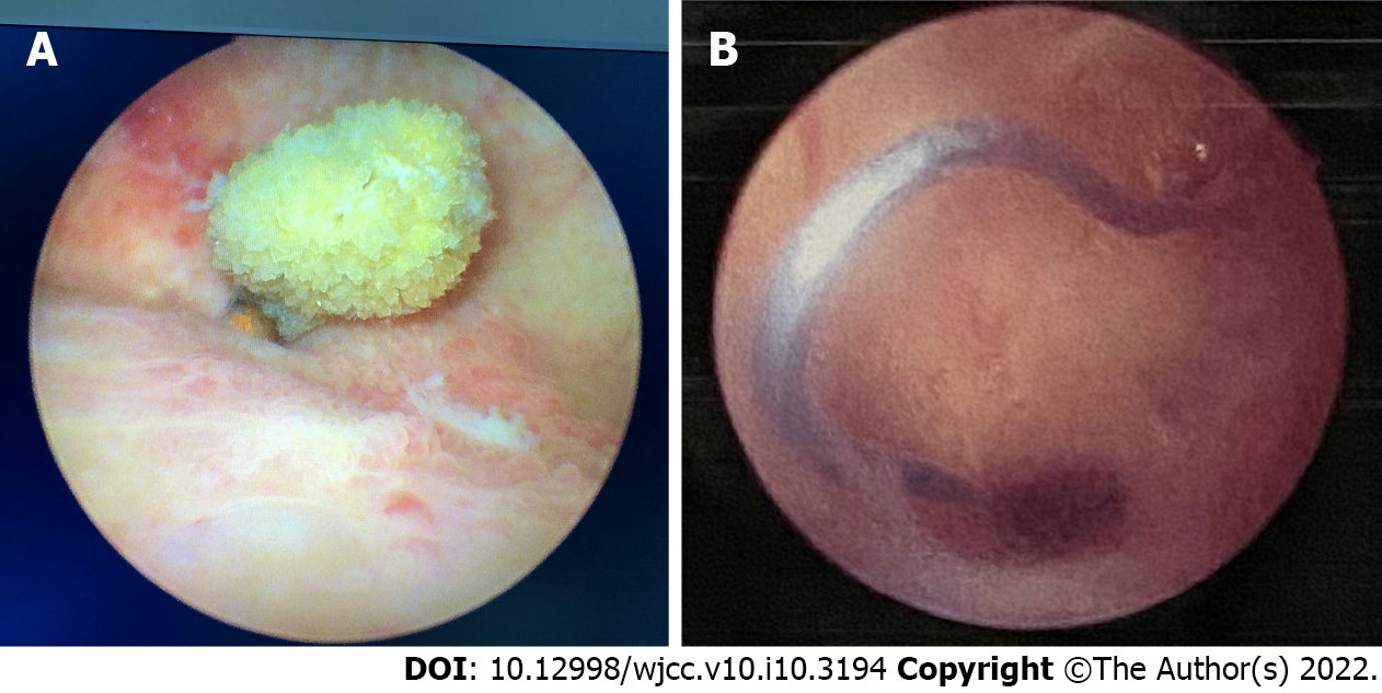
Figure 2 Cystoscopy and hysteroscopic images of an ectopic intrauterine device.
A: Cystoscopy reveals an intravesical device with many attached stones embedded in the bladder wall; B: Hysteroscopy demonstrates a V-type intrauterine device embedded in the myometrium of the anterior wall of the cervical canal.
- Citation: Yu HT, Chen Y, Xie YP, Gan TB, Gou X. Ectopic intrauterine device in the bladder causing cystolithiasis: A case report. World J Clin Cases 2022; 10(10): 3194-3199
- URL: https://www.wjgnet.com/2307-8960/full/v10/i10/3194.htm
- DOI: https://dx.doi.org/10.12998/wjcc.v10.i10.3194


