Copyright
©The Author(s) 2022.
World J Clin Cases. Apr 6, 2022; 10(10): 3164-3169
Published online Apr 6, 2022. doi: 10.12998/wjcc.v10.i10.3164
Published online Apr 6, 2022. doi: 10.12998/wjcc.v10.i10.3164
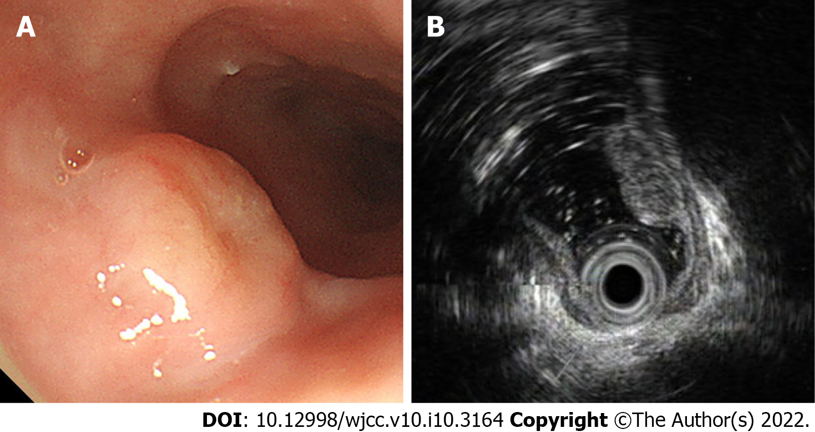
Figure 1 Endoscopic images of the tumor.
A: Gastroscopy demonstrated a disc-shaped protruding lesion of about 18 mm × 18 mm in size in the upper esophagus; B: Endoscopic ultrasonography demonstrated that the bulged lesion was highly echoic and homogeneous, originating from the muscularis mucosa.
- Citation: Tang N, Feng Z. Endoscopic submucosal dissection combined with adjuvant chemotherapy for early-stage neuroendocrine carcinoma of the esophagus: A case report. World J Clin Cases 2022; 10(10): 3164-3169
- URL: https://www.wjgnet.com/2307-8960/full/v10/i10/3164.htm
- DOI: https://dx.doi.org/10.12998/wjcc.v10.i10.3164


