Copyright
©The Author(s) 2022.
World J Clin Cases. Apr 6, 2022; 10(10): 3156-3163
Published online Apr 6, 2022. doi: 10.12998/wjcc.v10.i10.3156
Published online Apr 6, 2022. doi: 10.12998/wjcc.v10.i10.3156
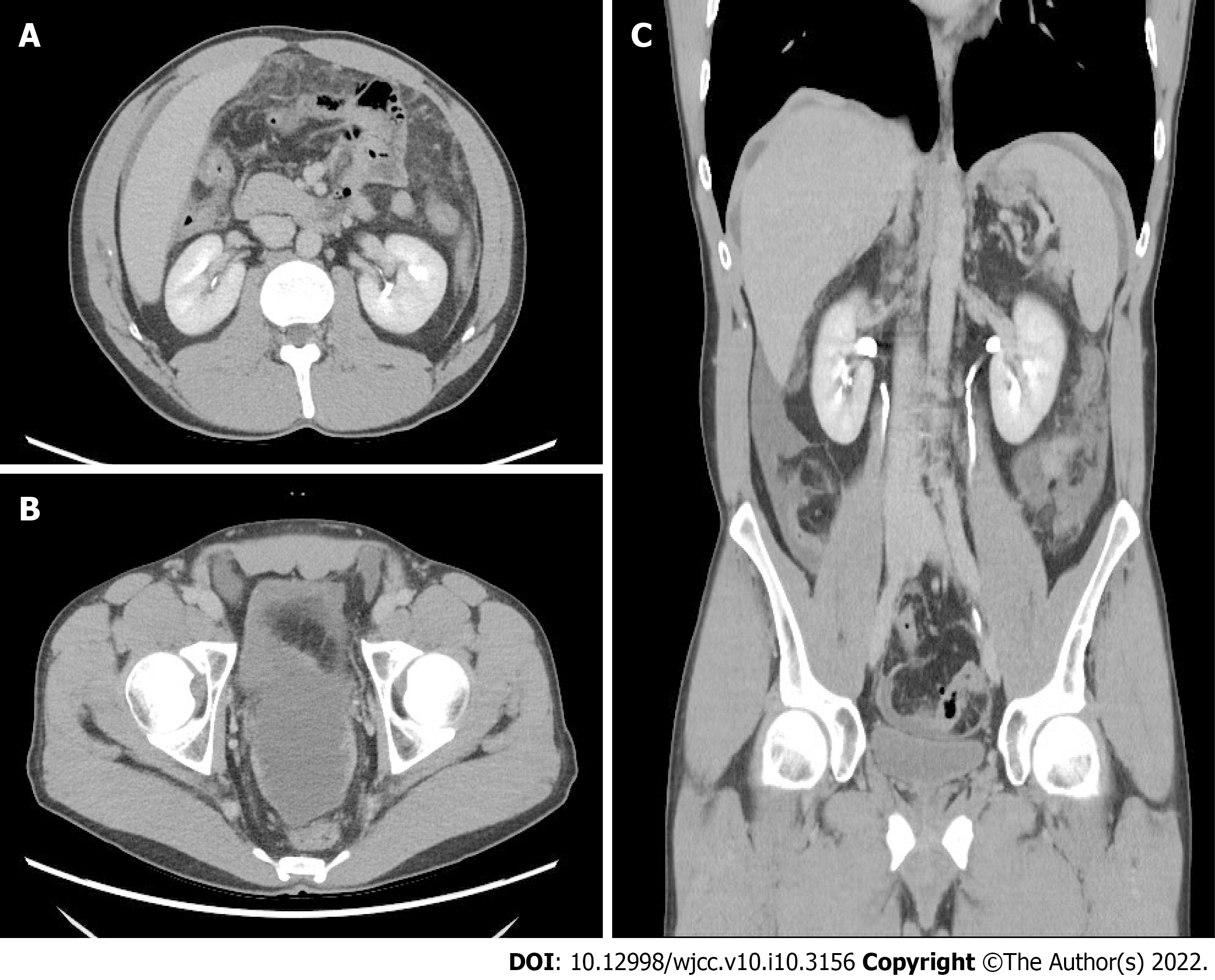
Figure 1 Abdominal contrast-enhanced computed tomography.
A: Axial view, contrast-enhanced computed tomography (CT) showed diffuse fat stranding infiltration within the greater omentum; B: Axial view, thickening of the pelvic peritoneum was enhanced by contrast material; C: Coronal view of the contrast-enhanced CT showed irregular thickening of the perihepatic peritoneum and minimal ascites over the bilateral subphrenic spaces, paracolic gutter, and pelvic cavity.
- Citation: Lin LC, Kuan WY, Shiu BH, Wang YT, Chao WR, Wang CC. Primary malignant peritoneal mesothelioma mimicking tuberculous peritonitis: A case report. World J Clin Cases 2022; 10(10): 3156-3163
- URL: https://www.wjgnet.com/2307-8960/full/v10/i10/3156.htm
- DOI: https://dx.doi.org/10.12998/wjcc.v10.i10.3156


