Copyright
©The Author(s) 2022.
World J Clin Cases. Jan 7, 2022; 10(1): 62-70
Published online Jan 7, 2022. doi: 10.12998/wjcc.v10.i1.62
Published online Jan 7, 2022. doi: 10.12998/wjcc.v10.i1.62
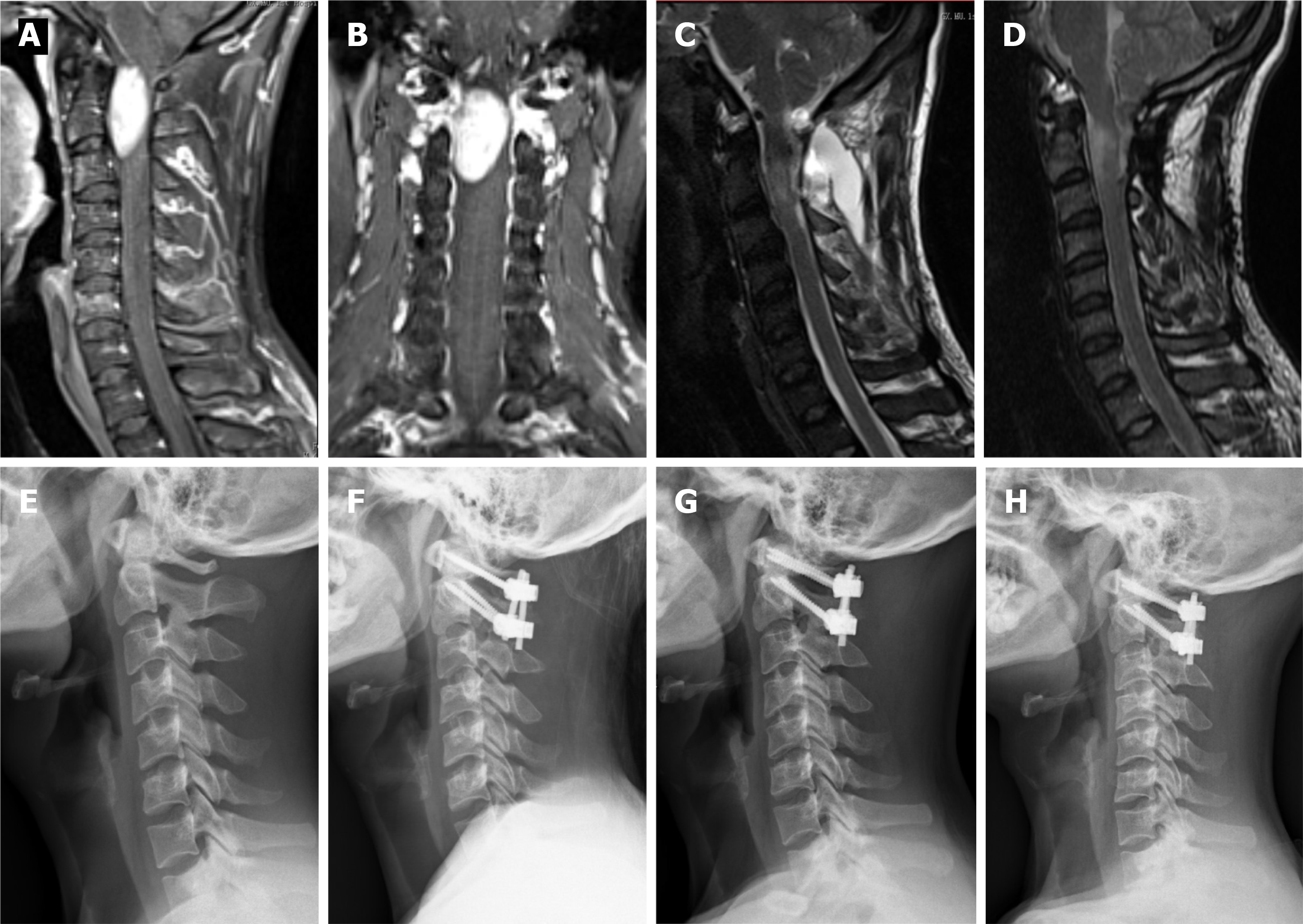
Figure 1 This 29-year-old male patient experienced a significant deterioration in his neurological condition and was unable to walk after 3 mo (Case 2).
A: Preoperative sagittal enhanced magnetic resonance imaging (MRI): an intradural extramedullary tumor located anterior to the atlantoaxial spinal cord; B: Preoperative coronal enhanced MRI: an intradural extramedullary tumor extending into the spinal canal at C1-C2 Levels; C, D: 3 mo and 31 mo postoperative sagittal T2-weighted MRI showing complete tumor resection; E: Preoperative X-ray: cervical sagittal alignment shows a loss of cervical lordosis; F, G, H: 3 mo, 18 mo and 31 mo postoperative X-ray: C1-C2 fixation with cervical pedicle screws and titanium plates.
- Citation: Meng DH, Wang JQ, Yang KX, Chen WY, Pan C, Jiang H. Surgical resection of intradural extramedullary tumors in the atlantoaxial spine via a posterior approach. World J Clin Cases 2022; 10(1): 62-70
- URL: https://www.wjgnet.com/2307-8960/full/v10/i1/62.htm
- DOI: https://dx.doi.org/10.12998/wjcc.v10.i1.62


