Copyright
©The Author(s) 2022.
World J Clin Cases. Jan 7, 2022; 10(1): 388-396
Published online Jan 7, 2022. doi: 10.12998/wjcc.v10.i1.388
Published online Jan 7, 2022. doi: 10.12998/wjcc.v10.i1.388
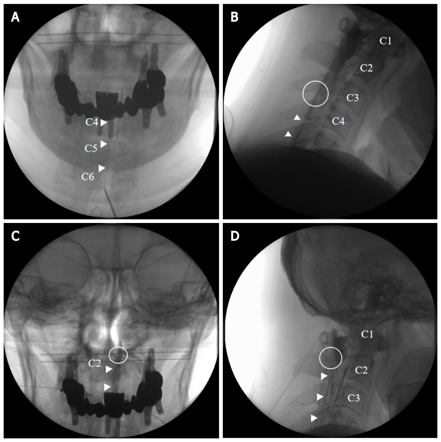
Figure 4 C-arm imaging.
A and B: Anteroposterior and lateral views showing an epidurogram during initial targeted cervical epidural blood patch; C and D: Anteroposterior and lateral views showing an epidurogram during repeat targeted cervical epidural blood patch. Arrowhead: Catheter; Circle: Catheter tip.
- Citation: Choi SH, Lee YY, Kim WJ. Epidural blood patch for spontaneous intracranial hypotension with subdural hematoma: A case report and review of literature. World J Clin Cases 2022; 10(1): 388-396
- URL: https://www.wjgnet.com/2307-8960/full/v10/i1/388.htm
- DOI: https://dx.doi.org/10.12998/wjcc.v10.i1.388


