Copyright
©The Author(s) 2022.
World J Clin Cases. Jan 7, 2022; 10(1): 388-396
Published online Jan 7, 2022. doi: 10.12998/wjcc.v10.i1.388
Published online Jan 7, 2022. doi: 10.12998/wjcc.v10.i1.388
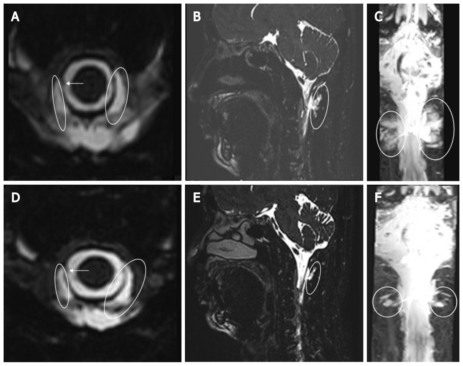
Figure 3 Magnetic resonance myelography.
A-C: Axial, sagittal, and coronal scanning images showing focal dural sac defect at the right C1/2 level and cerebrospinal fluid (CSF) collection in bilateral and posterior C1/2 epidural space; D-F: Axial, sagittal, and coronal scanning images showing focal dural sac thinning at the right C1/2 level and decreased size of CSF accumulation. White arrow: Dural sac defect; Oval: CSF accumulation.
- Citation: Choi SH, Lee YY, Kim WJ. Epidural blood patch for spontaneous intracranial hypotension with subdural hematoma: A case report and review of literature. World J Clin Cases 2022; 10(1): 388-396
- URL: https://www.wjgnet.com/2307-8960/full/v10/i1/388.htm
- DOI: https://dx.doi.org/10.12998/wjcc.v10.i1.388


