Copyright
©The Author(s) 2022.
World J Clin Cases. Jan 7, 2022; 10(1): 345-352
Published online Jan 7, 2022. doi: 10.12998/wjcc.v10.i1.345
Published online Jan 7, 2022. doi: 10.12998/wjcc.v10.i1.345
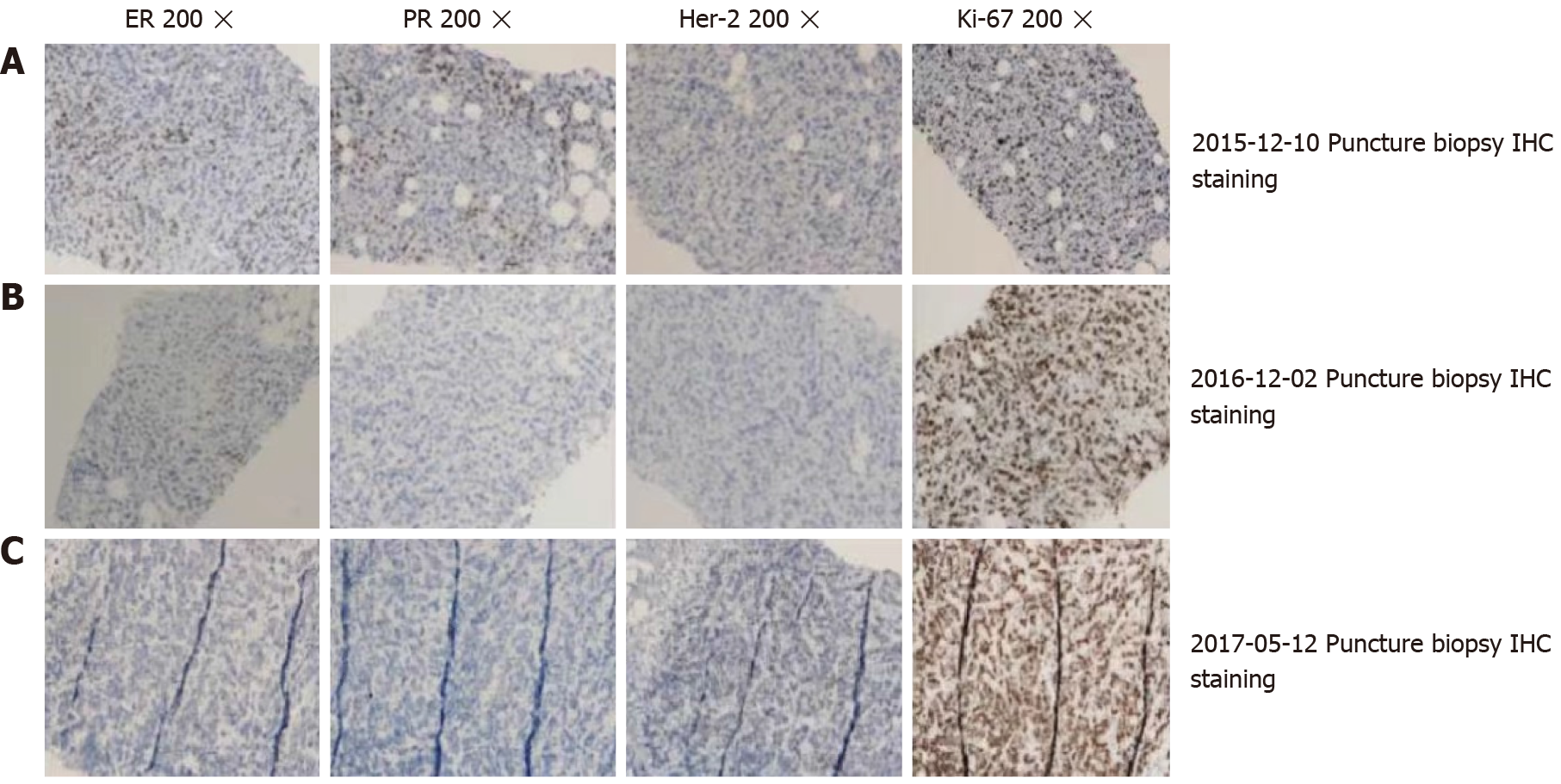
Figure 3 Immunohistochemical results at different puncture time points.
A: Immunohistochemical staining of the breast puncture biopsy (20 × magnification); B: High expression of ER (+), PR (+), and Ki-67 (40%), and low expression of Her-2 (-); C: Triple negative expression of ER (-), PR (-), and HER2 (-), and high expression of Ki-67 (60%); D: Low expression of ER (10%+), PR (-), HER2 (-), and high expression of Ki-67 (90%). The pathology results in A, B, and C were obtained from the same patient at different time points of puncture.
- Citation: Li ZH, Wang F, Zhang P, Xue P, Zhu SJ. Diagnosis and guidance of treatment of breast cancer cutaneous metastases by multiple needle biopsy: A case report. World J Clin Cases 2022; 10(1): 345-352
- URL: https://www.wjgnet.com/2307-8960/full/v10/i1/345.htm
- DOI: https://dx.doi.org/10.12998/wjcc.v10.i1.345


