Copyright
©The Author(s) 2022.
World J Clin Cases. Jan 7, 2022; 10(1): 331-337
Published online Jan 7, 2022. doi: 10.12998/wjcc.v10.i1.331
Published online Jan 7, 2022. doi: 10.12998/wjcc.v10.i1.331
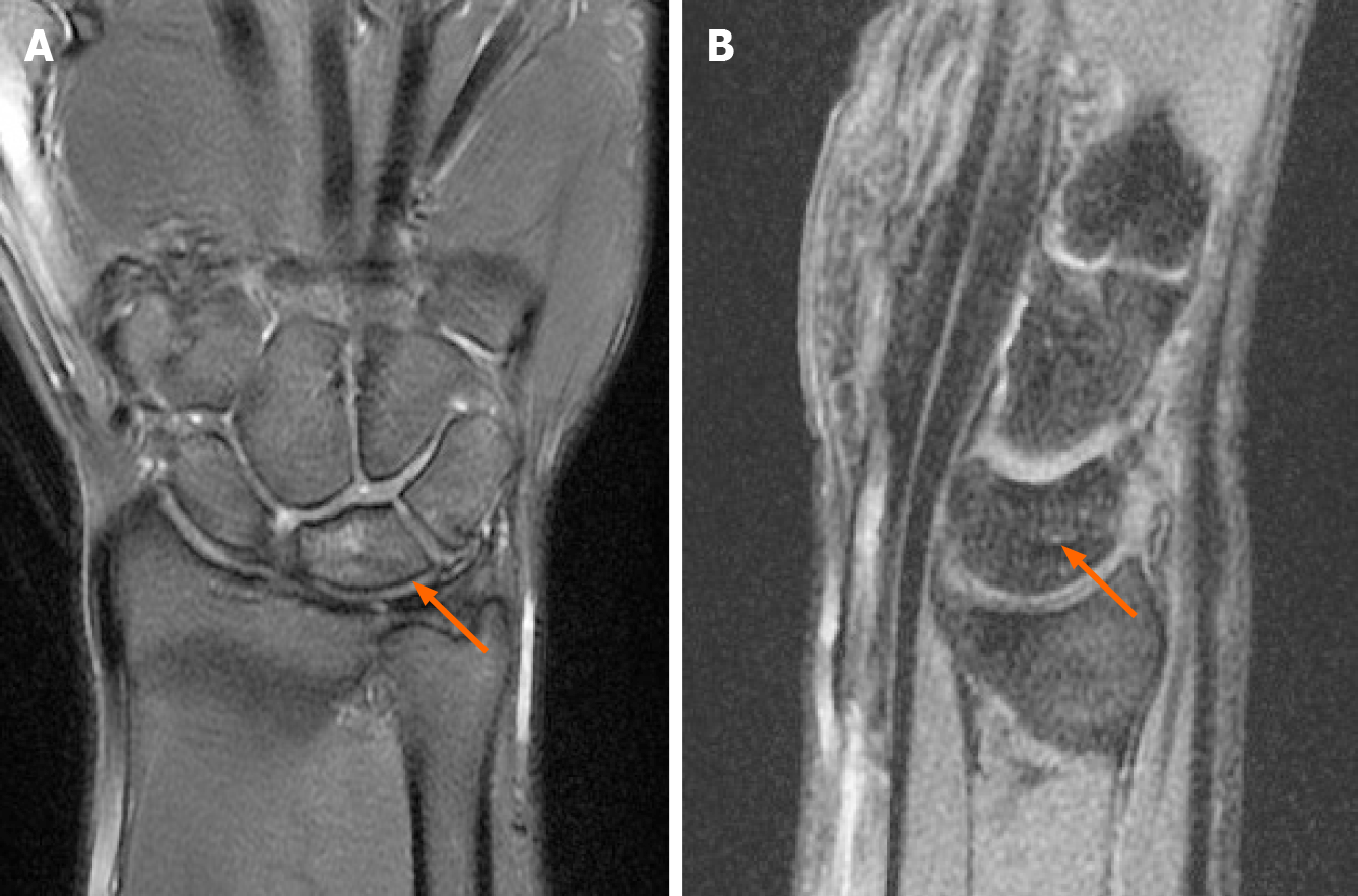
Figure 7 Magnetic resonance imaging follow-up 18 mo postoperatively.
A: Coronal section; B: Sagittal section. These findings showed almost completely resorption of bone edema of the lunate (arrow) and did not progress into osteonecrosis.
- Citation: Li LY, Lin CJ, Ko CY. Lunate dislocation with avulsed triquetral fracture: A case report. World J Clin Cases 2022; 10(1): 331-337
- URL: https://www.wjgnet.com/2307-8960/full/v10/i1/331.htm
- DOI: https://dx.doi.org/10.12998/wjcc.v10.i1.331


