Copyright
©The Author(s) 2022.
World J Clin Cases. Jan 7, 2022; 10(1): 316-322
Published online Jan 7, 2022. doi: 10.12998/wjcc.v10.i1.316
Published online Jan 7, 2022. doi: 10.12998/wjcc.v10.i1.316
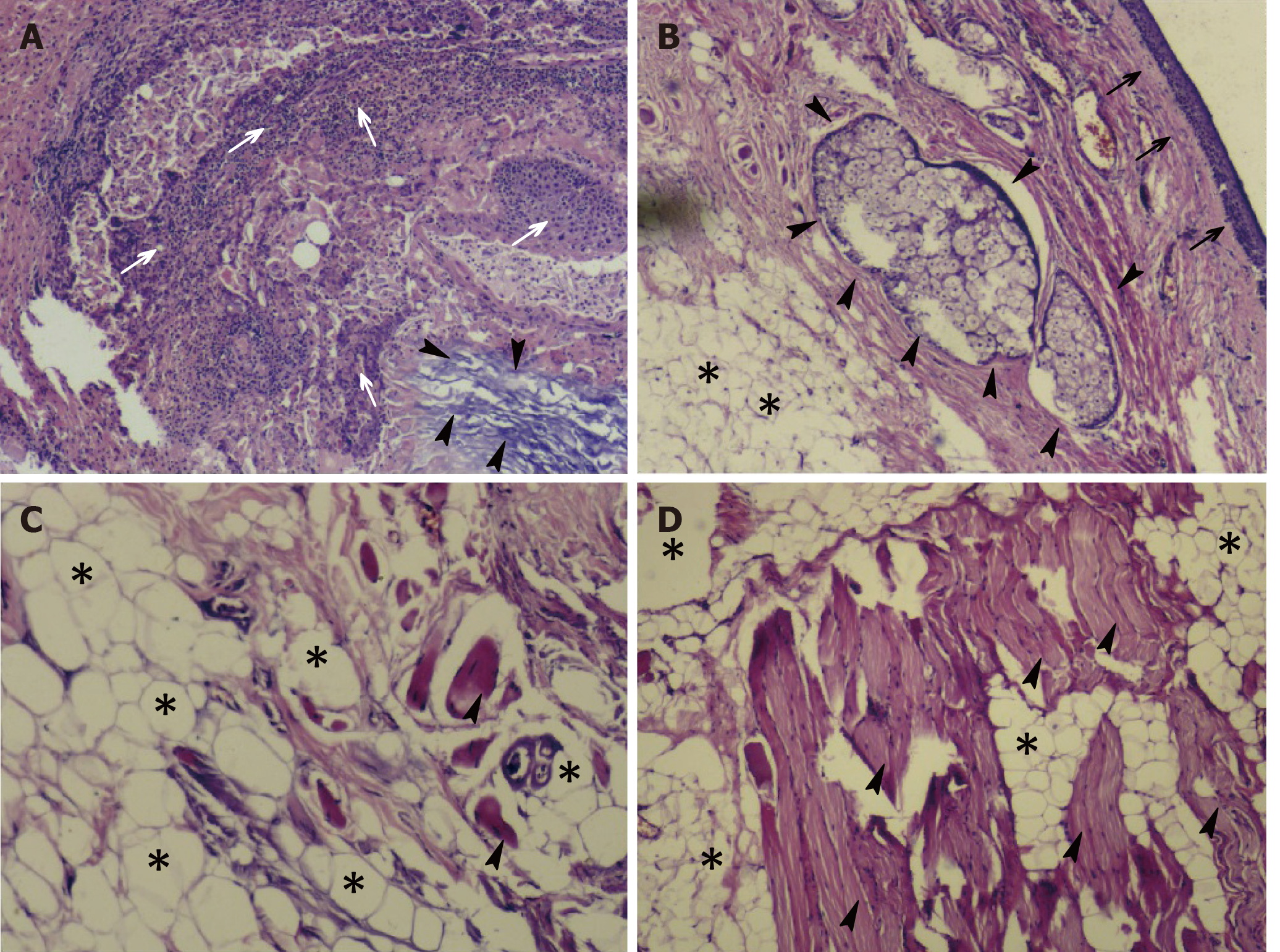
Figure 4 Histopathological appearance of the patient’s resected tumor mass.
A: Photomicrograph of the eustachian tube teratoma shows a mass with keratinized squamous epithelium (arrowheads) and chronic inflammatory cells (white arrows); B: Squamous epithelium (black arrows), adipose tissue (*) and sebaceous glands (arrowheads); C and D: Adipose tissue (*) and mature skeletal muscle tissue (arrowheads). All images are original magnification of × 100.
- Citation: Li JY, Sun LX, Hu N, Song GS, Dou WQ, Gong RZ, Li CT. Eustachian tube teratoma: A case report. World J Clin Cases 2022; 10(1): 316-322
- URL: https://www.wjgnet.com/2307-8960/full/v10/i1/316.htm
- DOI: https://dx.doi.org/10.12998/wjcc.v10.i1.316


