Copyright
©The Author(s) 2022.
World J Clin Cases. Jan 7, 2022; 10(1): 316-322
Published online Jan 7, 2022. doi: 10.12998/wjcc.v10.i1.316
Published online Jan 7, 2022. doi: 10.12998/wjcc.v10.i1.316
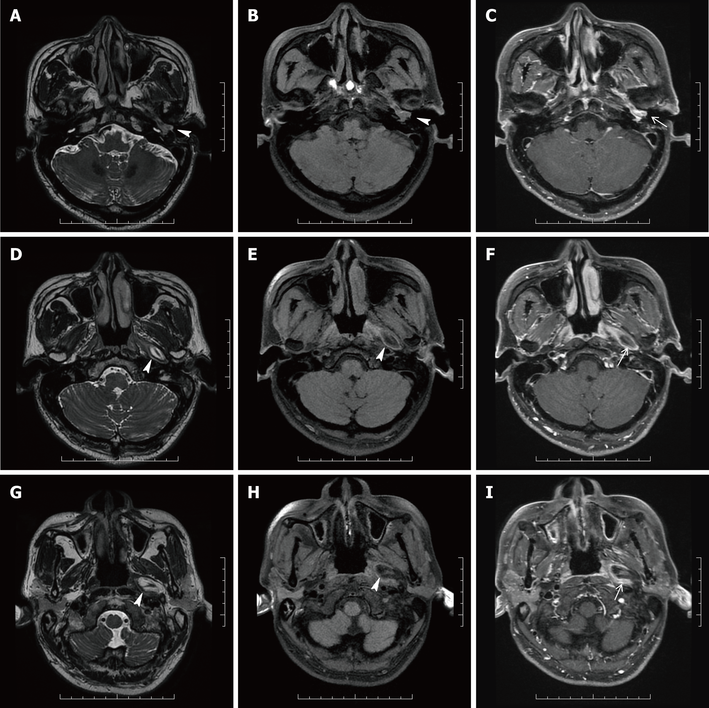
Figure 3 Magnetic resonance imaging of the patient’s head and neck.
A, D, G: Three-dimensional (3D) T2 weighted image (WI); B, E, H: Fat suppression (FS) 3D T1WI; C, F, I: FS 3D T1WI with contrast (C+); A-C: Magnetic resonance (MR) images in the transverse plane showed the part of the mass which was a homogeneous lesion with slightly higher signal intensity and with enhancement (white arrowheads) in the tympanum and external auditory canal; D-F, G-I: MR images in the transverse plane showed the part of the mass which was a well-defined, homogeneous lesion with high signal intensity along the left eustachian tube (white arrowheads), and on FS 3D T1WI, a lesion with decreased signal intensity consistent with macroscopic fat and with a contrast-enhancing rim (black arrowhead) was seen.
- Citation: Li JY, Sun LX, Hu N, Song GS, Dou WQ, Gong RZ, Li CT. Eustachian tube teratoma: A case report. World J Clin Cases 2022; 10(1): 316-322
- URL: https://www.wjgnet.com/2307-8960/full/v10/i1/316.htm
- DOI: https://dx.doi.org/10.12998/wjcc.v10.i1.316


