Copyright
©The Author(s) 2022.
World J Clin Cases. Jan 7, 2022; 10(1): 283-288
Published online Jan 7, 2022. doi: 10.12998/wjcc.v10.i1.283
Published online Jan 7, 2022. doi: 10.12998/wjcc.v10.i1.283
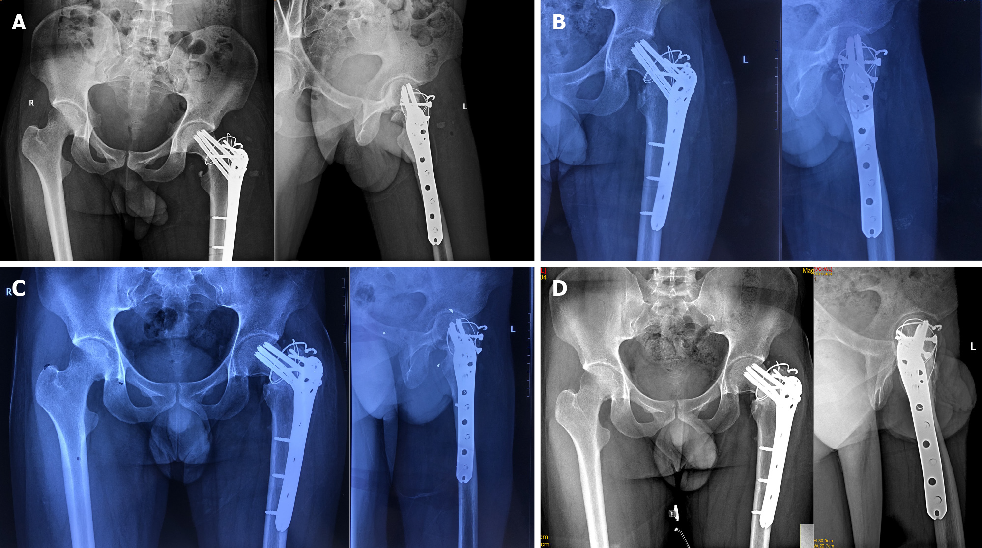
Figure 2 Radiographs after surgery.
A: The postoperative X-rays showed that the fractures were well reduced, the cervico-diaphyseal angle of the femur was satisfactory, and the internal fixation position was normal; B: The X-rays of 3 mo after surgery showed that the fracture lines were blurred; C: The X-rays of 12 mo after surgery showed that the fractured end of the femoral neck had collapsed, the cervico-diaphyseal angle had reduced, and the fracture lines were blurred; D: The X-rays of 24 mo were similar to those at 12 mo postoperatively, and the hip varus deformity did not significantly worsen.
- Citation: Li ZY, Cheng WD, Qi L, Yu SS, Jing JH. Complex proximal femoral fracture in a young patient followed up for 3 years: A case report. World J Clin Cases 2022; 10(1): 283-288
- URL: https://www.wjgnet.com/2307-8960/full/v10/i1/283.htm
- DOI: https://dx.doi.org/10.12998/wjcc.v10.i1.283


