Copyright
©The Author(s) 2022.
World J Clin Cases. Jan 7, 2022; 10(1): 275-282
Published online Jan 7, 2022. doi: 10.12998/wjcc.v10.i1.275
Published online Jan 7, 2022. doi: 10.12998/wjcc.v10.i1.275
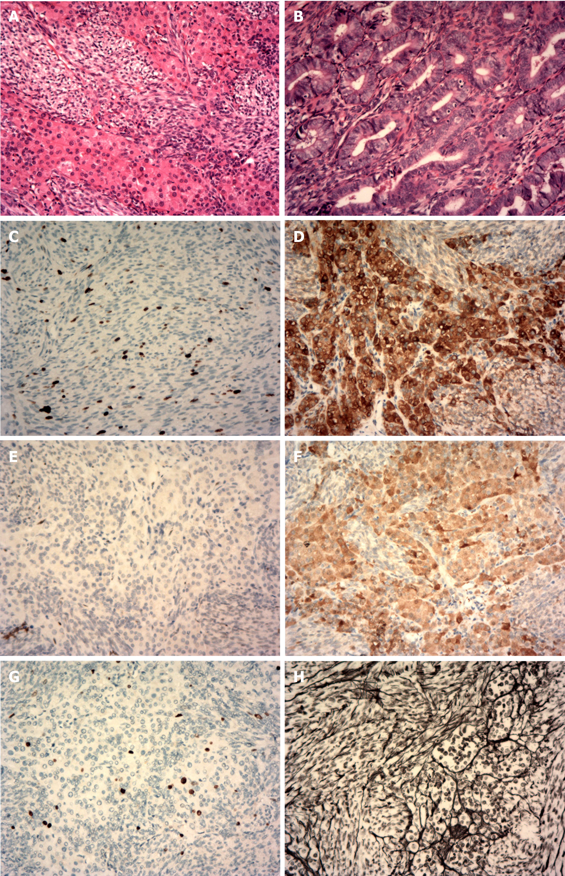
Figure 3 Histopathological and immunohistochemical staining findings.
A: The tumor cells of the right ovarian lesion were spindle-shaped and bundle-like with sheet-like arrangement, and the cells were densely arranged without obvious atypia. Eosinophilic cell nests with rich cytoplasm were visible in the focal area; B: The densely arranged endometrial glands were hyperplastic, and some glandular lumens were irregular; C: Cytokeratin (focal+); D: Inhibin (part+); E: Wilm’s tumor protein (WT1, part+); F: Calretinin (+); G: Ki-67 (approximately 10% +); H: Net staining showed mostly surrounding single cells.
- Citation: Wang J, Yang Q, Zhang NN, Wang DD. Recurrent postmenopausal bleeding - just endometrial disease or ovarian sex cord-stromal tumor? A case report. World J Clin Cases 2022; 10(1): 275-282
- URL: https://www.wjgnet.com/2307-8960/full/v10/i1/275.htm
- DOI: https://dx.doi.org/10.12998/wjcc.v10.i1.275


