Copyright
©The Author(s) 2022.
World J Clin Cases. Jan 7, 2022; 10(1): 275-282
Published online Jan 7, 2022. doi: 10.12998/wjcc.v10.i1.275
Published online Jan 7, 2022. doi: 10.12998/wjcc.v10.i1.275
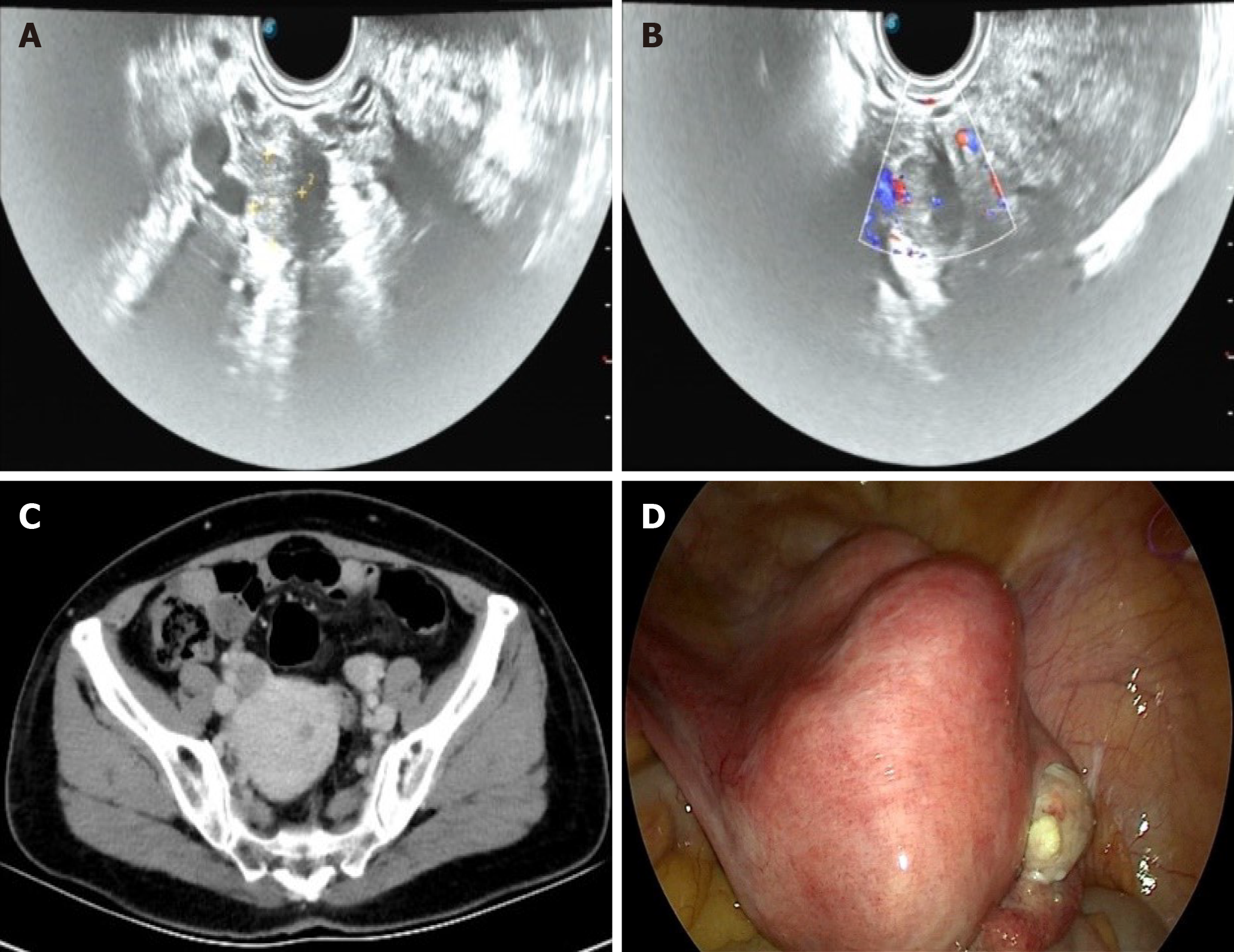
Figure 2 Imaging findings of the patient before and during the fifth operation.
A, B: Transvaginal ultrasonography: A 1.6 cm × 1.2 cm × 1.2 cm mass with hypoechoic was seen in the right ovary, the boundary was clear. Blood-flow signals were detected with color Doppler flow imaging; C: Abdominal computed tomography: There was a low-density nodule with a size of approximately 1.5 cm × 1.0 cm in the right ovary, with clear borders; D: Laparoscopy: The uterus was obviously large and irregular, with multiple fibroids nodules. A yellow protruding lesion of approximately 1 cm was observed on the surface of the right ovary.
- Citation: Wang J, Yang Q, Zhang NN, Wang DD. Recurrent postmenopausal bleeding - just endometrial disease or ovarian sex cord-stromal tumor? A case report. World J Clin Cases 2022; 10(1): 275-282
- URL: https://www.wjgnet.com/2307-8960/full/v10/i1/275.htm
- DOI: https://dx.doi.org/10.12998/wjcc.v10.i1.275


