Copyright
©2013 Baishideng Publishing Group Co.
World J Clin Cases. Dec 16, 2013; 1(9): 285-289
Published online Dec 16, 2013. doi: 10.12998/wjcc.v1.i9.285
Published online Dec 16, 2013. doi: 10.12998/wjcc.v1.i9.285
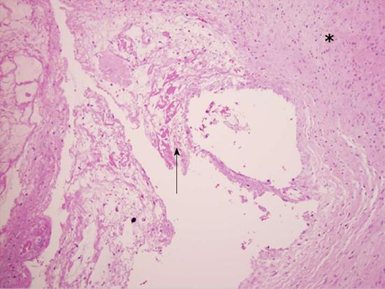
Figure 2 Case 2, pathological findings of midline ganglion cyst (hematoxylin eosin staining, × 400).
Photomicrograph (original magnification × 400) reveals proteinaceous material (arrow) surrounded by dense fibro-connective tissue (asterisk), without the presence of synovial epithelium. These findings confirm the diagnosis of a ganglion cyst.
- Citation: Pindrik J, Macki M, Bydon M, Maleki Z, Bydon A. Midline synovial and ganglion cysts causing neurogenic claudication. World J Clin Cases 2013; 1(9): 285-289
- URL: https://www.wjgnet.com/2307-8960/full/v1/i9/285.htm
- DOI: https://dx.doi.org/10.12998/wjcc.v1.i9.285


