Copyright
©2013 Baishideng Publishing Group Co.
World J Clin Cases. Dec 16, 2013; 1(9): 285-289
Published online Dec 16, 2013. doi: 10.12998/wjcc.v1.i9.285
Published online Dec 16, 2013. doi: 10.12998/wjcc.v1.i9.285
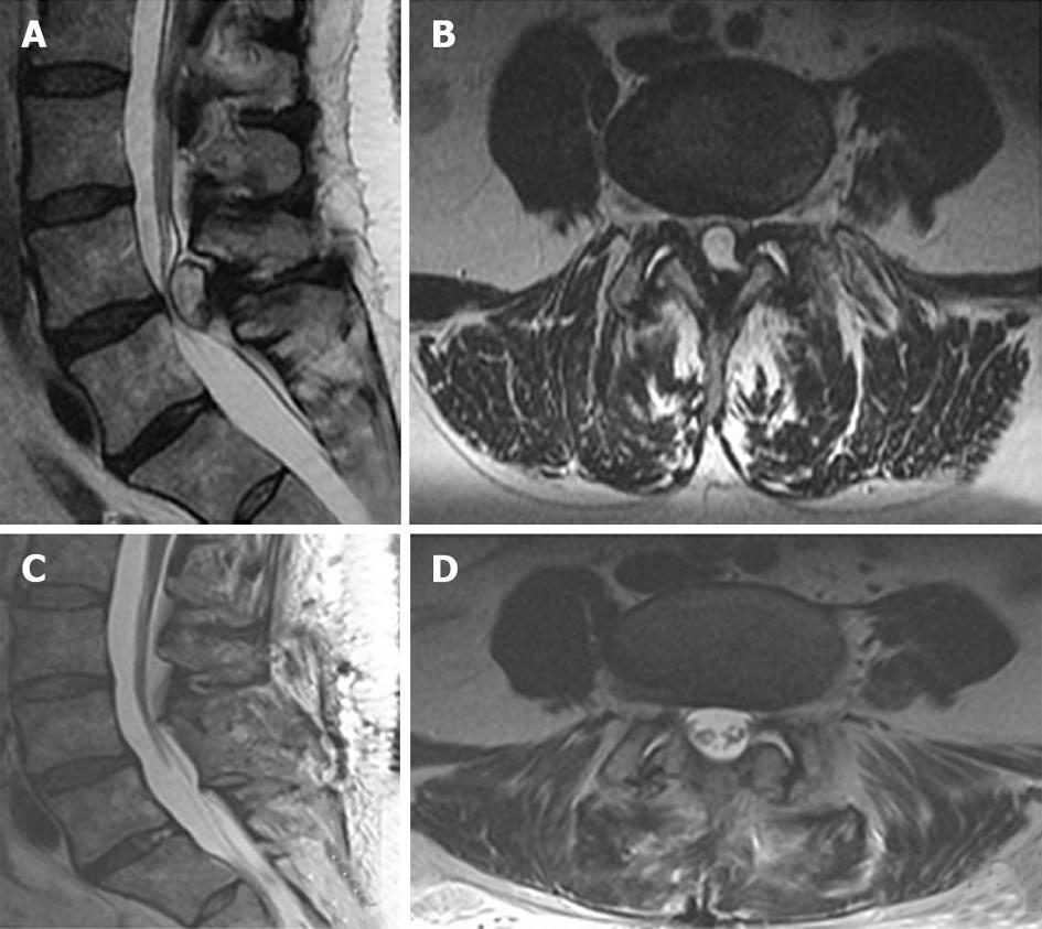
Figure 1 T2-weighted imaging of case 2.
A, B: In the preoperative images, sagittal (A) and axial (B) T2 weighted magnetic resonance imaging displays a large intraspinal cystic lesion at the L4-5 level causing stenosis and thecal sac compression. The cystic lesion appears to have an eccentric component extending towards the left facet joint; C, D: Post-operative imaging following decompressive laminectomy and cyst excision. Bilateral laminectomies at L4-5 and cyst resection provide adequate decompression of the dorsal thecal sac and cauda equina, as shown in sagittal (C) and axial (D) images.
- Citation: Pindrik J, Macki M, Bydon M, Maleki Z, Bydon A. Midline synovial and ganglion cysts causing neurogenic claudication. World J Clin Cases 2013; 1(9): 285-289
- URL: https://www.wjgnet.com/2307-8960/full/v1/i9/285.htm
- DOI: https://dx.doi.org/10.12998/wjcc.v1.i9.285


