Copyright
©2013 Baishideng Publishing Group Co.
World J Clin Cases. Dec 16, 2013; 1(9): 276-284
Published online Dec 16, 2013. doi: 10.12998/wjcc.v1.i9.276
Published online Dec 16, 2013. doi: 10.12998/wjcc.v1.i9.276
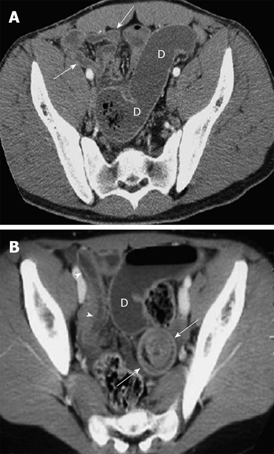
Figure 7 Meckel’s diverticulum.
A: Small-bowel obstruction shown on computed tomography (CT) in an 18-year-old boy with pathologically proven Meckel’s diverticulum; B: CT image in an 11-year-old girl shows intussusception (arrows) as a bowel loop containing alternating rings of attenuation. Note dilated proximal small bowel (D) and collapsed terminal ileum (arrowheads).
- Citation: Yang WC, Chen CY, Wu HP. Etiology of non-traumatic acute abdomen in pediatric emergency departments. World J Clin Cases 2013; 1(9): 276-284
- URL: https://www.wjgnet.com/2307-8960/full/v1/i9/276.htm
- DOI: https://dx.doi.org/10.12998/wjcc.v1.i9.276


