Copyright
©2013 Baishideng Publishing Group Co.
World J Clin Cases. Dec 16, 2013; 1(9): 276-284
Published online Dec 16, 2013. doi: 10.12998/wjcc.v1.i9.276
Published online Dec 16, 2013. doi: 10.12998/wjcc.v1.i9.276
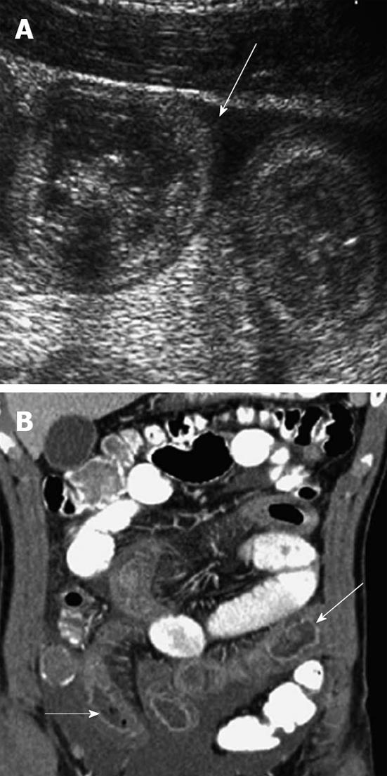
Figure 5 Henoch-Schonlein purpura.
A: Sagittal ultrasound image shows more moderate wall thickening of small bowel with ascites; B: Coronal computed tomography shows long segments of thickened, enhancing, fluid filled small bowel (arrows).
- Citation: Yang WC, Chen CY, Wu HP. Etiology of non-traumatic acute abdomen in pediatric emergency departments. World J Clin Cases 2013; 1(9): 276-284
- URL: https://www.wjgnet.com/2307-8960/full/v1/i9/276.htm
- DOI: https://dx.doi.org/10.12998/wjcc.v1.i9.276


