Copyright
©2013 Baishideng Publishing Group Co.
World J Clin Cases. Dec 16, 2013; 1(9): 262-275
Published online Dec 16, 2013. doi: 10.12998/wjcc.v1.i9.262
Published online Dec 16, 2013. doi: 10.12998/wjcc.v1.i9.262
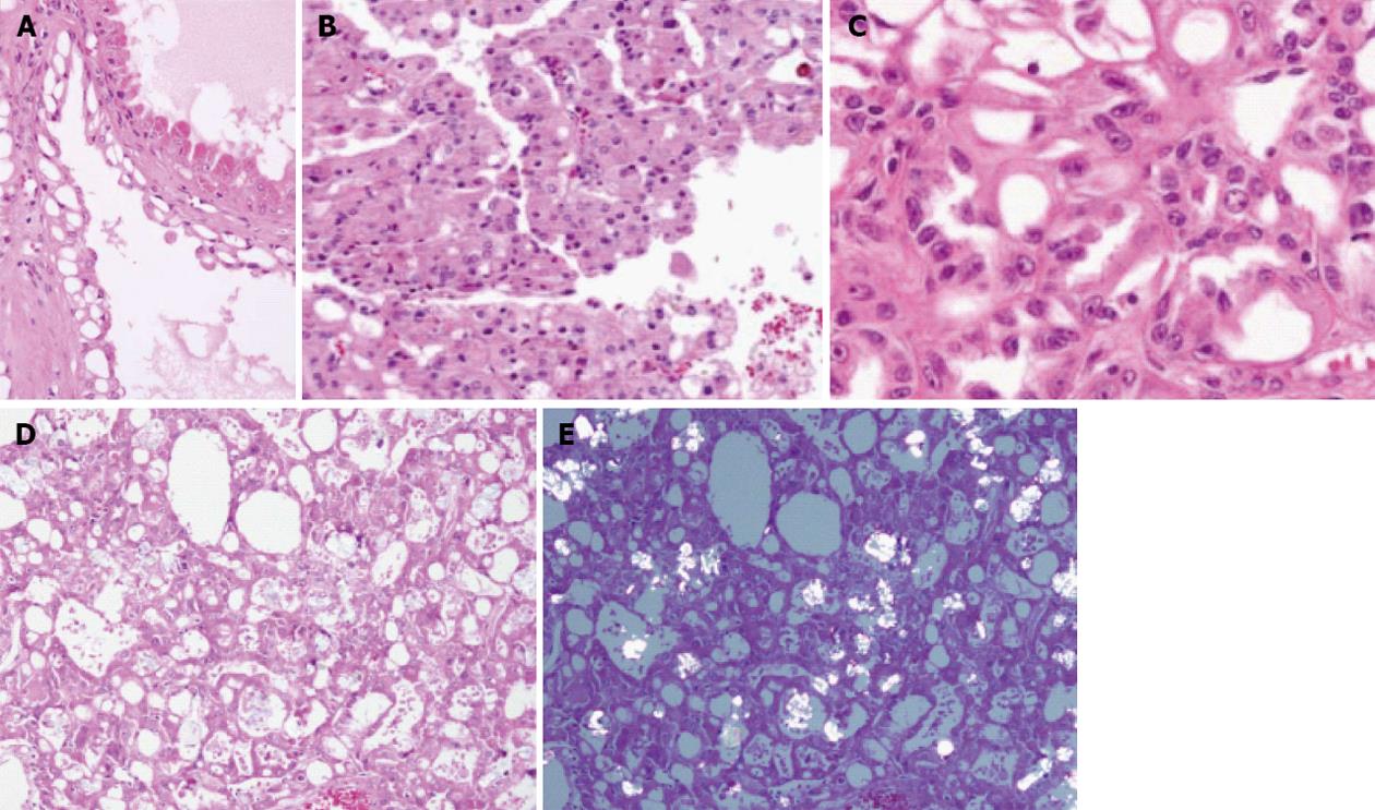
Figure 6 Acquired cystic disease-associated renal cell carcinoma.
A, B: The tumor cells in acquired cystic disease-associated renal cell carcinoma typically have abundant eosinophilic cytoplasm and are seen lining the cystic spaces (A, × 400), and sometimes also show a solid growth pattern (B, × 200); The tumor characteristically arranges in a sieve-like cribriform pattern; C: The nuclear features are consistent with a Fuhrman nuclear grade 3, with prominent nucleoli (× 600); D, E: Calcium oxalate crystals are often associated with these tumors, and can be seen on hematoxylin and eosin staining (D, × 600) and with polarization (E, × 100).
- Citation: Crumley SM, Divatia M, Truong L, Shen S, Ayala AG, Ro JY. Renal cell carcinoma: Evolving and emerging subtypes. World J Clin Cases 2013; 1(9): 262-275
- URL: https://www.wjgnet.com/2307-8960/full/v1/i9/262.htm
- DOI: https://dx.doi.org/10.12998/wjcc.v1.i9.262


