Copyright
©2013 Baishideng Publishing Group Co.
World J Clin Cases. Dec 16, 2013; 1(9): 262-275
Published online Dec 16, 2013. doi: 10.12998/wjcc.v1.i9.262
Published online Dec 16, 2013. doi: 10.12998/wjcc.v1.i9.262
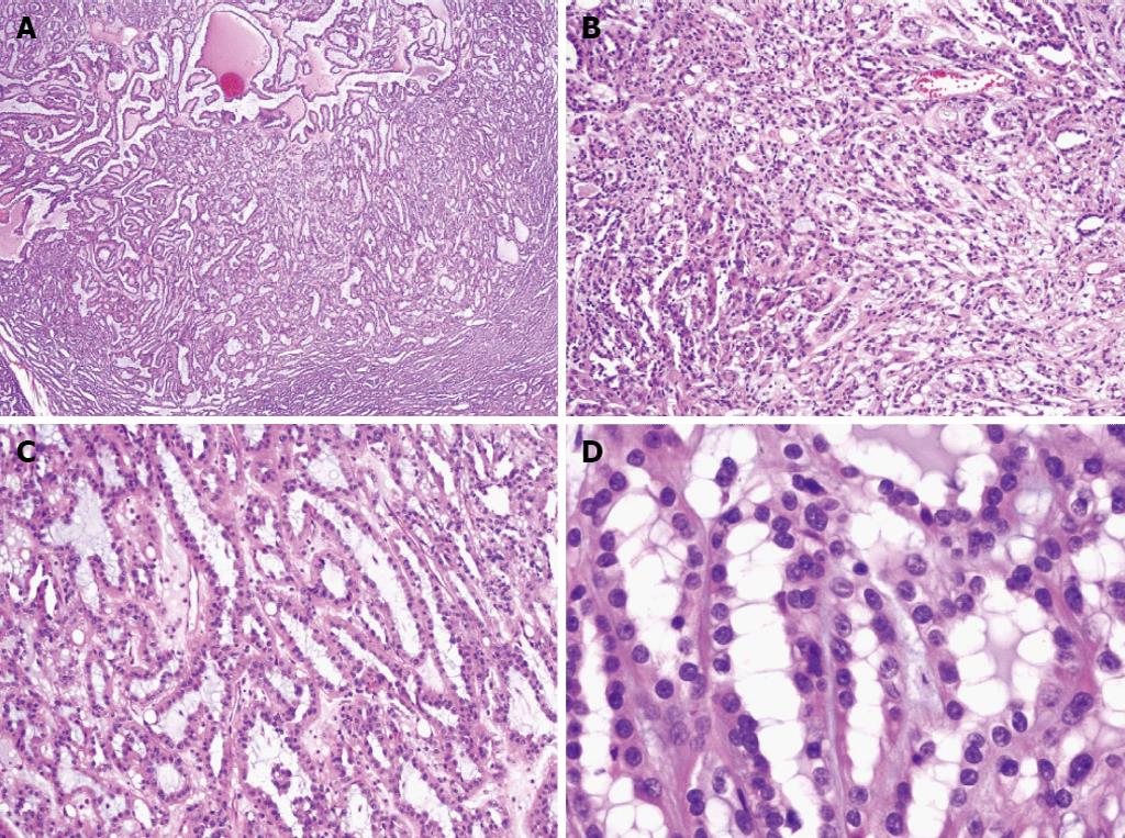
Figure 2 Mucinous and tubular spindle cell carcinoma (hematoxylin and eosin).
A: The tumor has a tubular pattern on low power with mucin present between glands (× 20); B: In some areas, a spindle cell pattern is also seen (× 40); C: The tubular pattern with intervening mucin is prominent in some tumors (× 100); D: Round nuclei with prominent nucleoli are evident on higher power (× 400).
- Citation: Crumley SM, Divatia M, Truong L, Shen S, Ayala AG, Ro JY. Renal cell carcinoma: Evolving and emerging subtypes. World J Clin Cases 2013; 1(9): 262-275
- URL: https://www.wjgnet.com/2307-8960/full/v1/i9/262.htm
- DOI: https://dx.doi.org/10.12998/wjcc.v1.i9.262


