Copyright
©2013 Baishideng Publishing Group Co.
World J Clin Cases. Oct 16, 2013; 1(7): 220-223
Published online Oct 16, 2013. doi: 10.12998/wjcc.v1.i7.220
Published online Oct 16, 2013. doi: 10.12998/wjcc.v1.i7.220
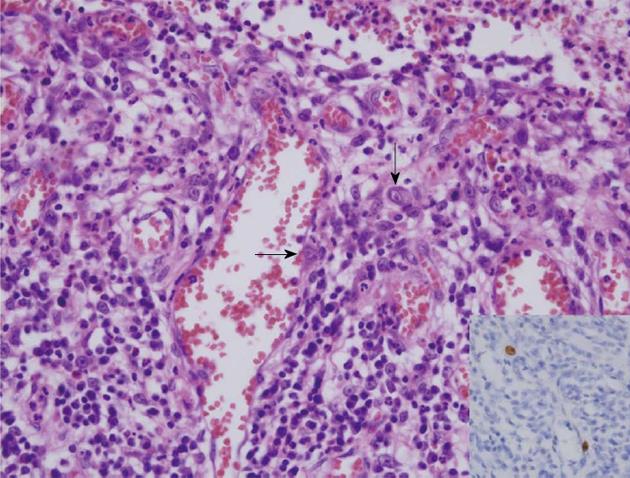
Figure 3 The ulcer bed was composed of granulation tissue with abundant vascular proliferation.
Many large atypical endothelial cells and stromal fibroblasts with the formation of intranuclear inclusion bodies were noted (arrows). These cells were positive for cytomegalovirus antibody (Inset).
- Citation: Jun YJ, Sim J, Ahn HI, Han H, Kim H, Yi K, Rehman A, Jang SM, Jang K, Paik SS. Cytomegalovirus enteritis with jejunal perforation in a patient with endometrial adenocarcinoma. World J Clin Cases 2013; 1(7): 220-223
- URL: https://www.wjgnet.com/2307-8960/full/v1/i7/220.htm
- DOI: https://dx.doi.org/10.12998/wjcc.v1.i7.220


