Copyright
©2013 Baishideng Publishing Group Co.
World J Clin Cases. Sep 16, 2013; 1(6): 197-201
Published online Sep 16, 2013. doi: 10.12998/wjcc.v1.i6.197
Published online Sep 16, 2013. doi: 10.12998/wjcc.v1.i6.197
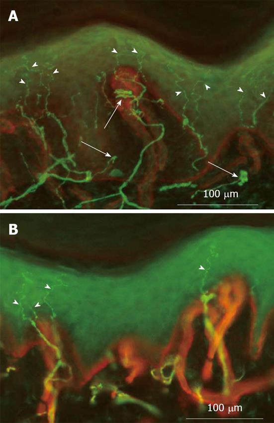
Figure 2 The figure illustrate the higher density of epidermal nerve fibres (arrowheads) in the allodynic skin (A), when compared to contralateral normal skin (B).
It also shows some abnormal patterns of nerve fibre regeneration (arrows) in the dermis of the allodynic skin. Indirect immunofluorescence method: in green, protein gene product 9.5 staining of nerve fibres; in red, type IV collagen staining of basement membrane and blood vessels.
- Citation: Buonocore M, Gagliano MC, Bonezzi C. Dynamic mechanical allodynia following finger amputation: Unexpected skin hyperinnervation. World J Clin Cases 2013; 1(6): 197-201
- URL: https://www.wjgnet.com/2307-8960/full/v1/i6/197.htm
- DOI: https://dx.doi.org/10.12998/wjcc.v1.i6.197


