Copyright
©2013 Baishideng Publishing Group Co.
World J Clin Cases. Jul 16, 2013; 1(4): 149-151
Published online Jul 16, 2013. doi: 10.12998/wjcc.v1.i4.149
Published online Jul 16, 2013. doi: 10.12998/wjcc.v1.i4.149
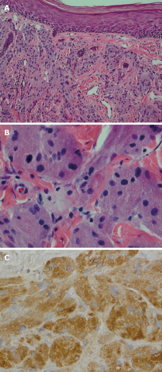
Figure 3 Photomicrograph.
A: clusters of nests of cells in lamina propria with squamous epithelium on the surface (HE, × 100); B: cells with granular eosinophilic cytoplasm. (HE, × 400); C: S-100 uptake, the brown color indicates the positive stain (S-100, × 400).
- Citation: Rivlin ME, Meeks GR, Ghafar MA, Lewin JR. Vulvar granular cell tumor. World J Clin Cases 2013; 1(4): 149-151
- URL: https://www.wjgnet.com/2307-8960/full/v1/i4/149.htm
- DOI: https://dx.doi.org/10.12998/wjcc.v1.i4.149


