Copyright
©2013 Baishideng Publishing Group Co.
World J Clin Cases. Jul 16, 2013; 1(4): 143-145
Published online Jul 16, 2013. doi: 10.12998/wjcc.v1.i4.143
Published online Jul 16, 2013. doi: 10.12998/wjcc.v1.i4.143
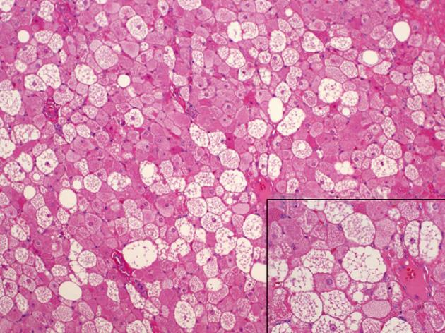
Figure 3 Microscopic evaluation of the hibernoma is characterized by vacuolated granular eosinophilic cells (hematoxylin-eosin, × 100).
The inset (hematoxylin-eosin, × 400) shows a high power view of granular and multivacuolated cells in hibernoma.
- Citation: Jaroszewski DE, De Petris G. Giant hibernoma of the thoracic pleura and chest wall. World J Clin Cases 2013; 1(4): 143-145
- URL: https://www.wjgnet.com/2307-8960/full/v1/i4/143.htm
- DOI: https://dx.doi.org/10.12998/wjcc.v1.i4.143


