Copyright
©2013 Baishideng.
World J Clin Cases. May 16, 2013; 1(2): 92-95
Published online May 16, 2013. doi: 10.12998/wjcc.v1.i2.92
Published online May 16, 2013. doi: 10.12998/wjcc.v1.i2.92
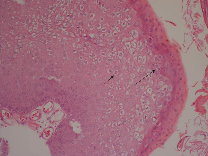
Figure 2 Histopathological findings (hematoxylin and eosin, × 200).
Histopathological examinations revealed the characteristic features of hyperkeratosis, papillomatosis, parakeratosis, acanthosis and koilocytosis. Koilocytosis indicates diseased cells caused by human papilloma virus infection. Characteristic squamous cells containing peri-nuclear clearing with condensation of peripheral cytoplasm are the typical pictures of koilocytic cells. Irregular, raisin-shaped nucleus (short arrow) and bi-nuclei cells (long arrow) may be present.
- Citation: Chang NC, Chien CY, Wu CC, Chai CY. Squamous papilloma in the external auditory canal: A common lesion in an uncommon site. World J Clin Cases 2013; 1(2): 92-95
- URL: https://www.wjgnet.com/2307-8960/full/v1/i2/92.htm
- DOI: https://dx.doi.org/10.12998/wjcc.v1.i2.92


