Copyright
©The Author(s) 2021.
World J Clin Cases. Nov 26, 2021; 9(33): 10315-10322
Published online Nov 26, 2021. doi: 10.12998/wjcc.v9.i33.10315
Published online Nov 26, 2021. doi: 10.12998/wjcc.v9.i33.10315
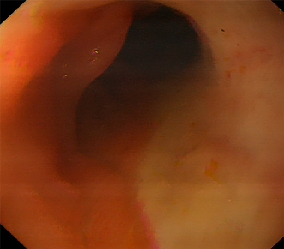
Figure 1
Esophagogastroduodenoscopy performed seven days before the acute gastrointestinal bleeding.
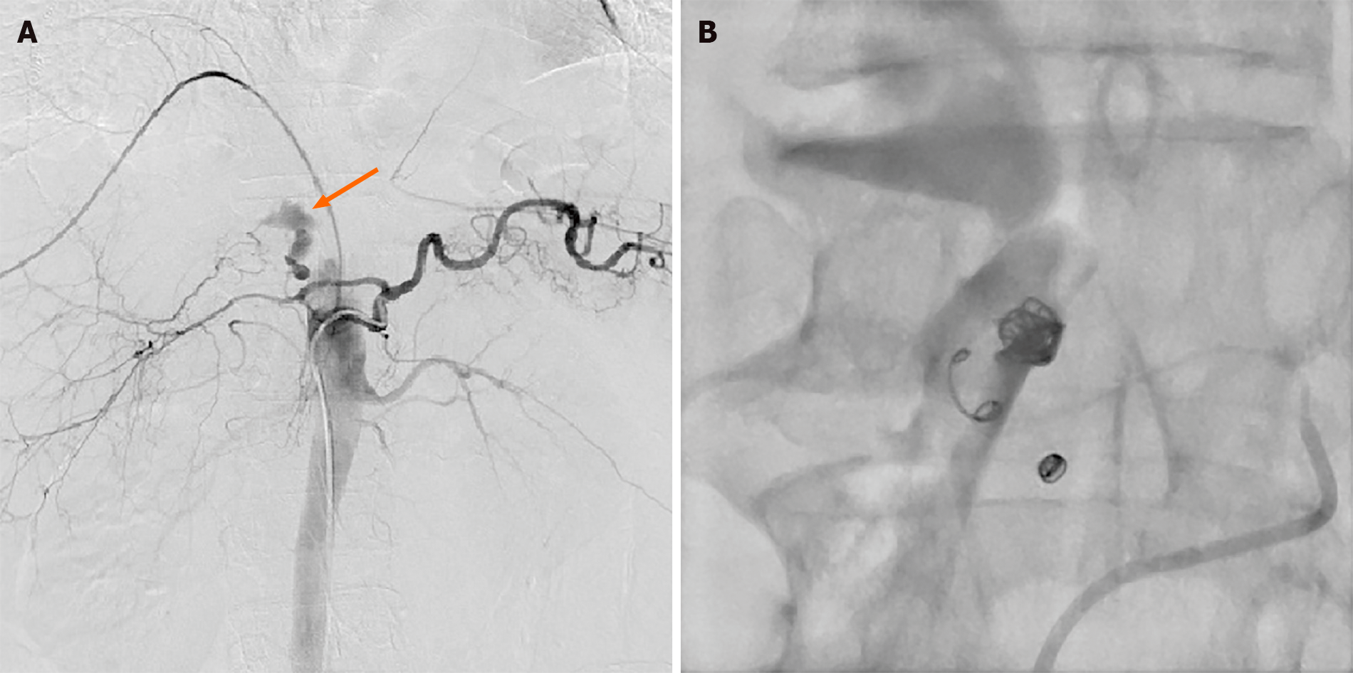
Figure 2 Digital subtraction angiography images before and after arterial embolization.
A: Digital subtraction angiography image showed extravasation of contrast agent at the branch of gastroduodenal artery (orange arrow); B: Digital subtraction angiography showed successful embolization of gastroduodenal artery branch.
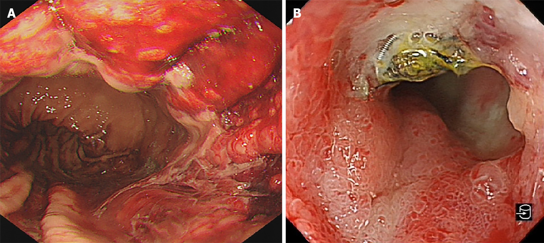
Figure 3 Esophagogastroduodenoscopy of the first hospitalization after interventional treatment and second hospitalization due to melena.
A: Esophagogastroduodenoscopy revealed diffuse congestion and erosion in the gastric corpus with bloody fluid; B: Esophagogastroduodenoscopy showed a duodenal ulcer caused by coil wiggle.
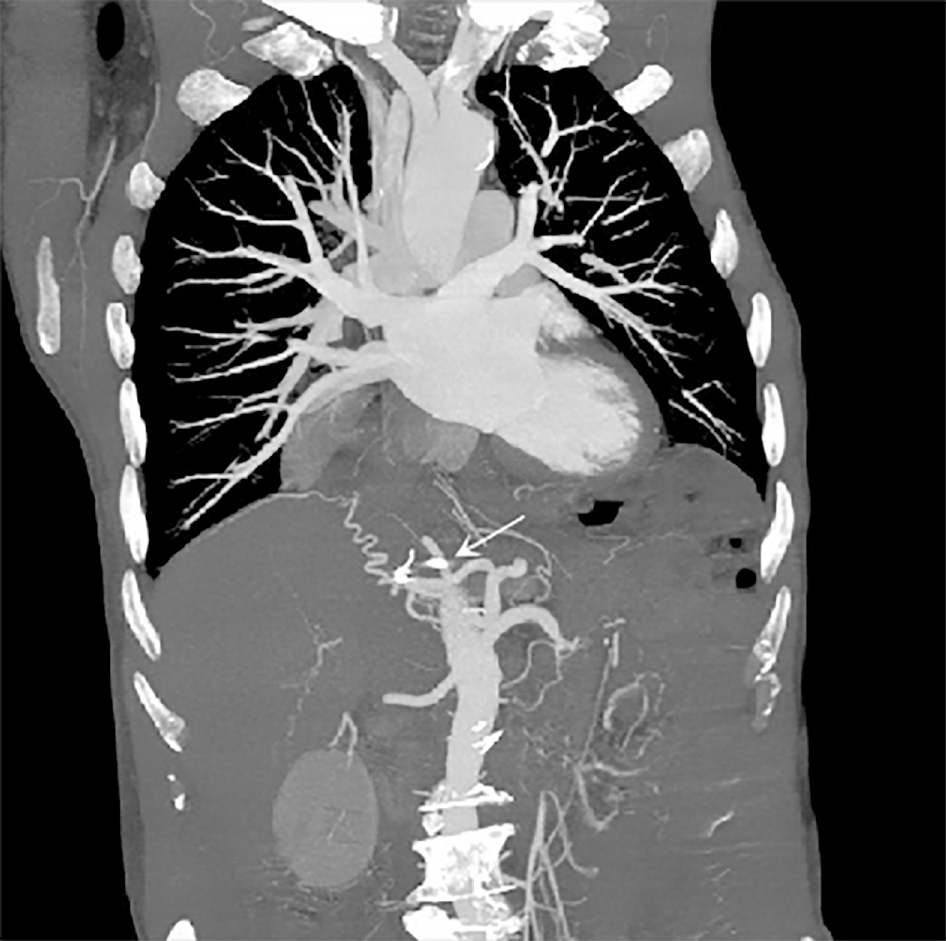
Figure 4
Abdominal computed tomography revealed the displaced coils.
- Citation: Xu S, Yang SX, Xue ZX, Xu CL, Cai ZZ, Xu CZ. Duodenal ulcer caused by coil wiggle after digital subtraction angiography-guided embolization: A case report. World J Clin Cases 2021; 9(33): 10315-10322
- URL: https://www.wjgnet.com/2307-8960/full/v9/i33/10315.htm
- DOI: https://dx.doi.org/10.12998/wjcc.v9.i33.10315


