Copyright
©The Author(s) 2020.
World J Clin Cases. Jul 6, 2020; 8(13): 2870-2875
Published online Jul 6, 2020. doi: 10.12998/wjcc.v8.i13.2870
Published online Jul 6, 2020. doi: 10.12998/wjcc.v8.i13.2870
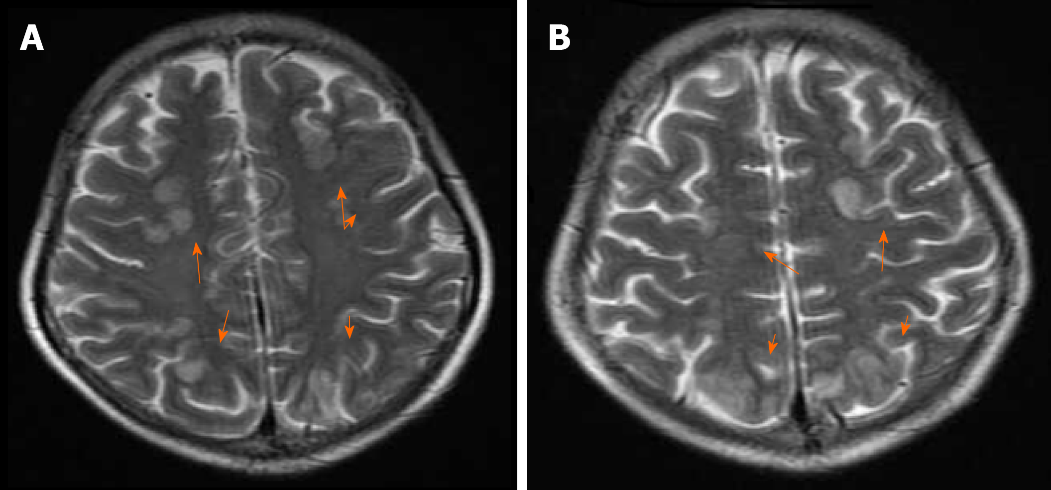
Figure 1 Initial brain magnetic resonance imaging findings (immediate post-seizure imaging).
A and B: A subcortical FLAIR hyperintensity area in the bilateral frontal, parietal, and occipital lobes (arrows), which was consistent with posterior reversible encephalopathy syndrome.
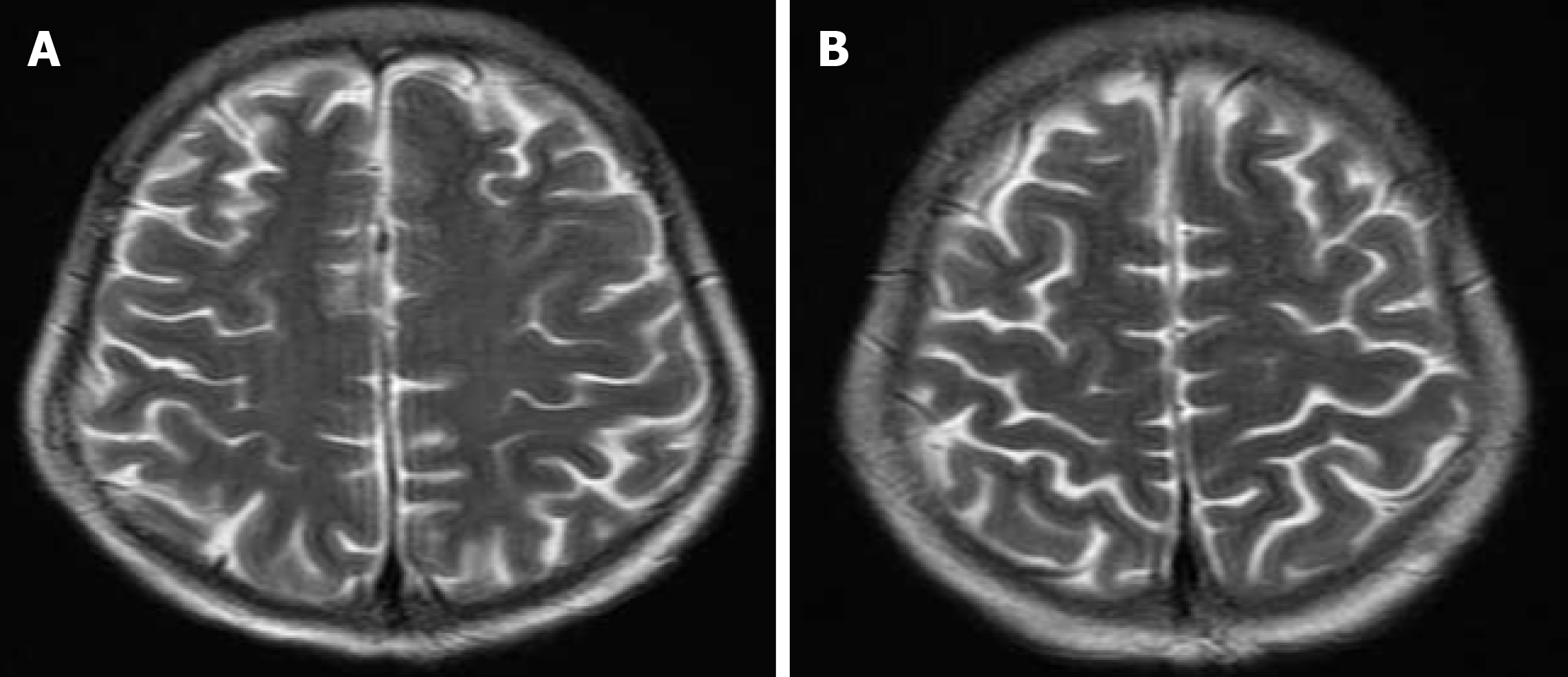
Figure 2 Follow-up brain magnetic resonance imaging findings (4 wk after switching tacrolimus to cyclosporine).
A and B: Resolution of the previous FLAIR hyperintensity signal abnormality.
- Citation: Liu JF, Shen T, Zhang YT. Posterior reversible encephalopathy syndrome and heart failure tacrolimus-induced after liver transplantation: A case report. World J Clin Cases 2020; 8(13): 2870-2875
- URL: https://www.wjgnet.com/2307-8960/full/v8/i13/2870.htm
- DOI: https://dx.doi.org/10.12998/wjcc.v8.i13.2870


