Copyright
©The Author(s) 2019.
World J Clin Cases. Nov 6, 2019; 7(21): 3649-3654
Published online Nov 6, 2019. doi: 10.12998/wjcc.v7.i21.3649
Published online Nov 6, 2019. doi: 10.12998/wjcc.v7.i21.3649
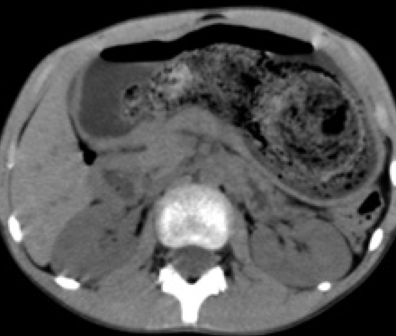
Figure 1 Axial computed tomography image showing a hairball in the stomach.
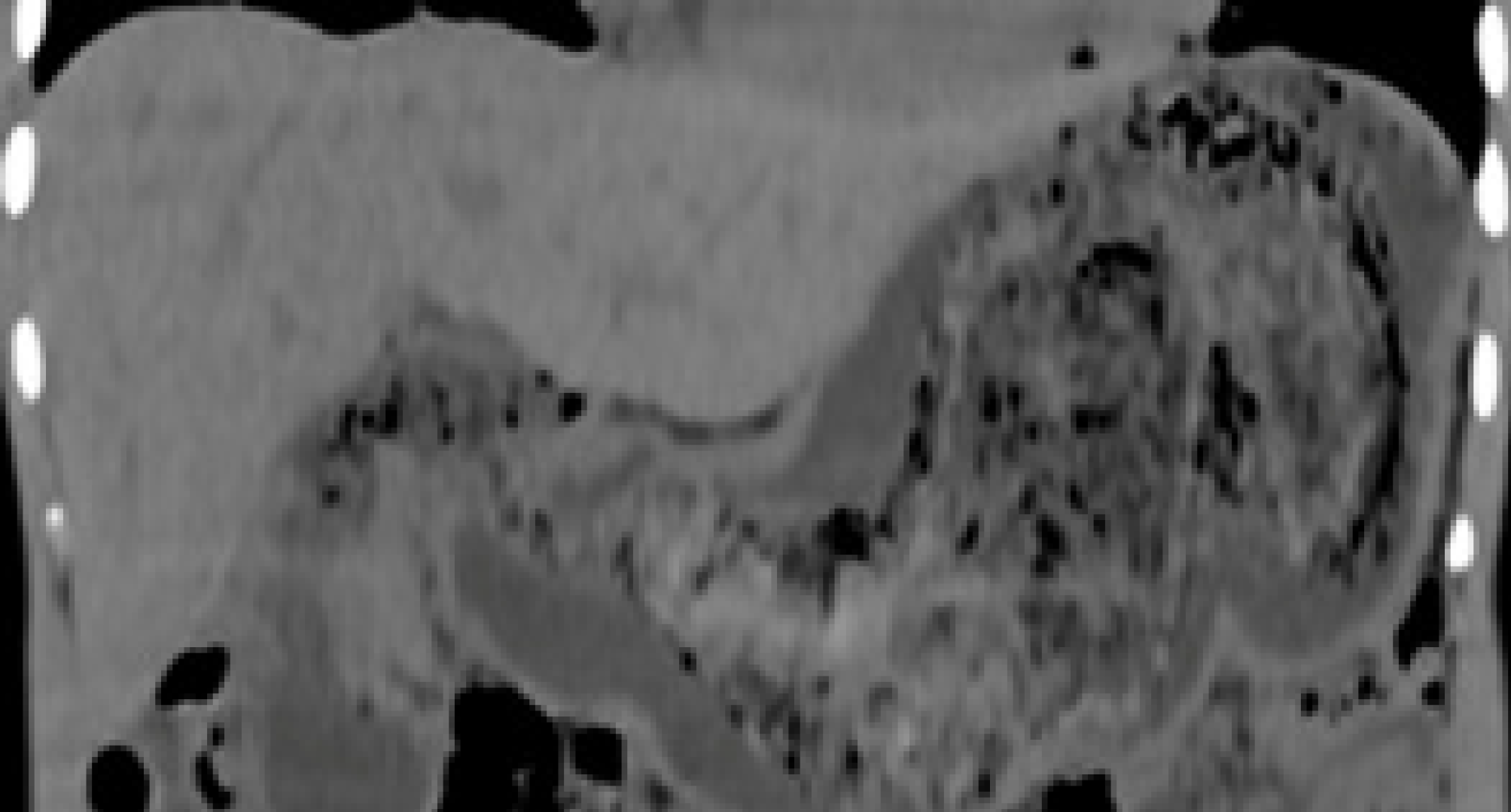
Figure 2 Coronary multiplanar reconstruction computed tomogrpahy image showing the extension of a hairball from the stomach into the second part of the duodenum.
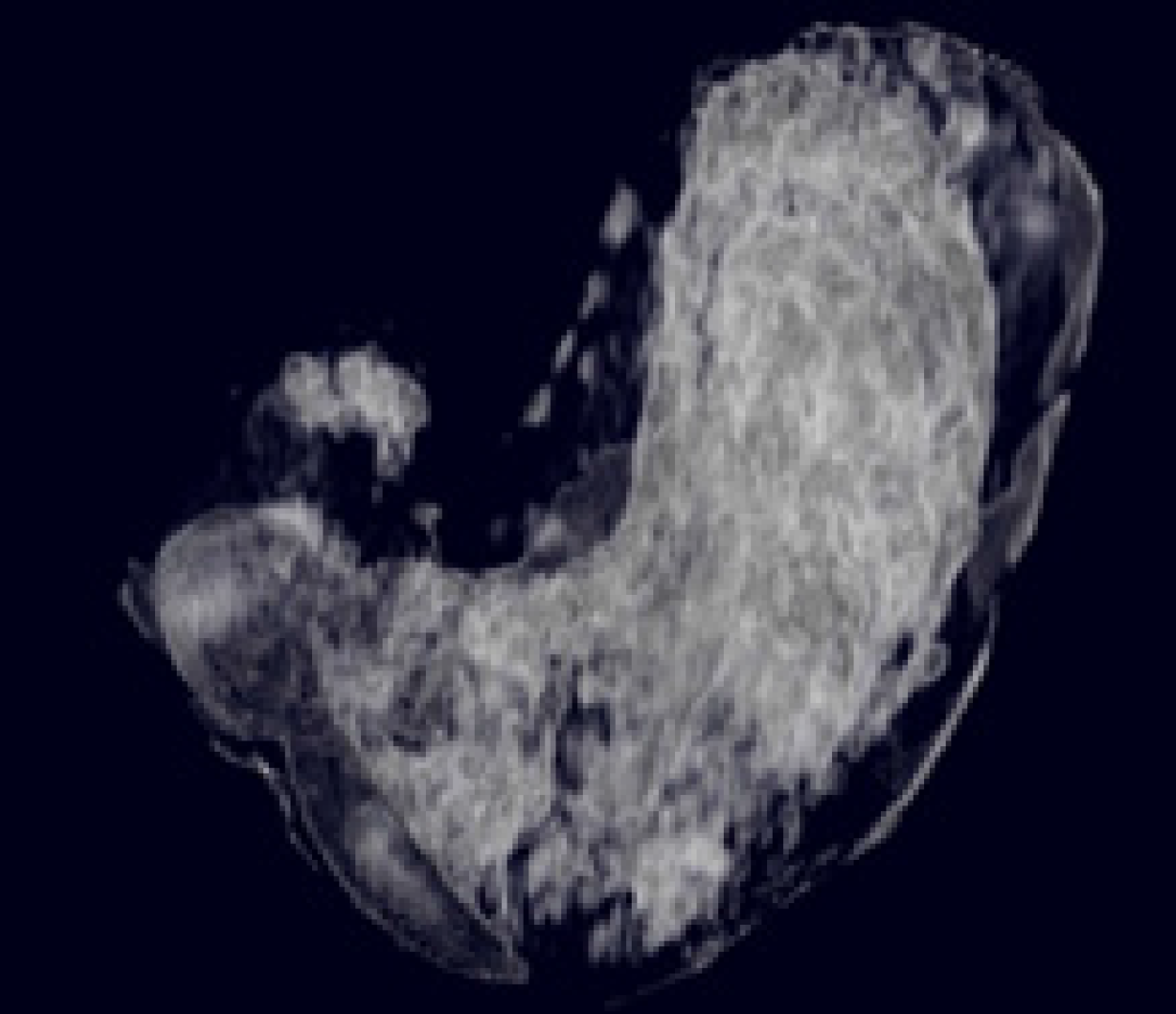
Figure 3 Three-dimension maximum intensity projection image showing the outline of a hairball defined by the air pockets in it.
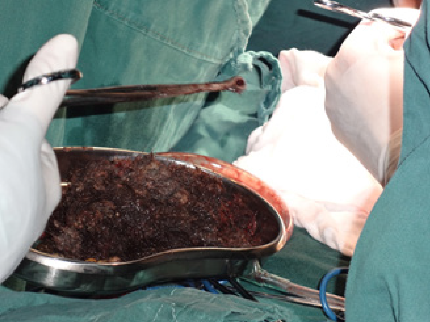
Figure 4 Postoperative photograph of the hair pulled from the stomach.
- Citation: Dong ZH, Yin F, Du SL, Mo ZH. Giant gastroduodenal trichobezoar: A case report. World J Clin Cases 2019; 7(21): 3649-3654
- URL: https://www.wjgnet.com/2307-8960/full/v7/i21/3649.htm
- DOI: https://dx.doi.org/10.12998/wjcc.v7.i21.3649


