Copyright
©The Author(s) 2019.
World J Clin Cases. Nov 6, 2019; 7(21): 3639-3648
Published online Nov 6, 2019. doi: 10.12998/wjcc.v7.i21.3639
Published online Nov 6, 2019. doi: 10.12998/wjcc.v7.i21.3639
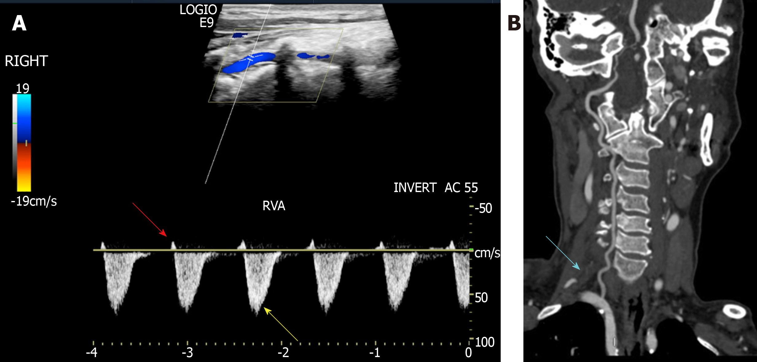
Figure 1 Imaging examinations.
A: Ultrasound showed that the right vertebral artery had a weak positive contractile peak in early systole (red arrow) followed by a large contractile wave in late systole below baseline (yellow arrow). Diastolic blood flow disappeared (type II right subclavian steal syndrome); B: Right vertebral artery dysplasia (blue arrow).
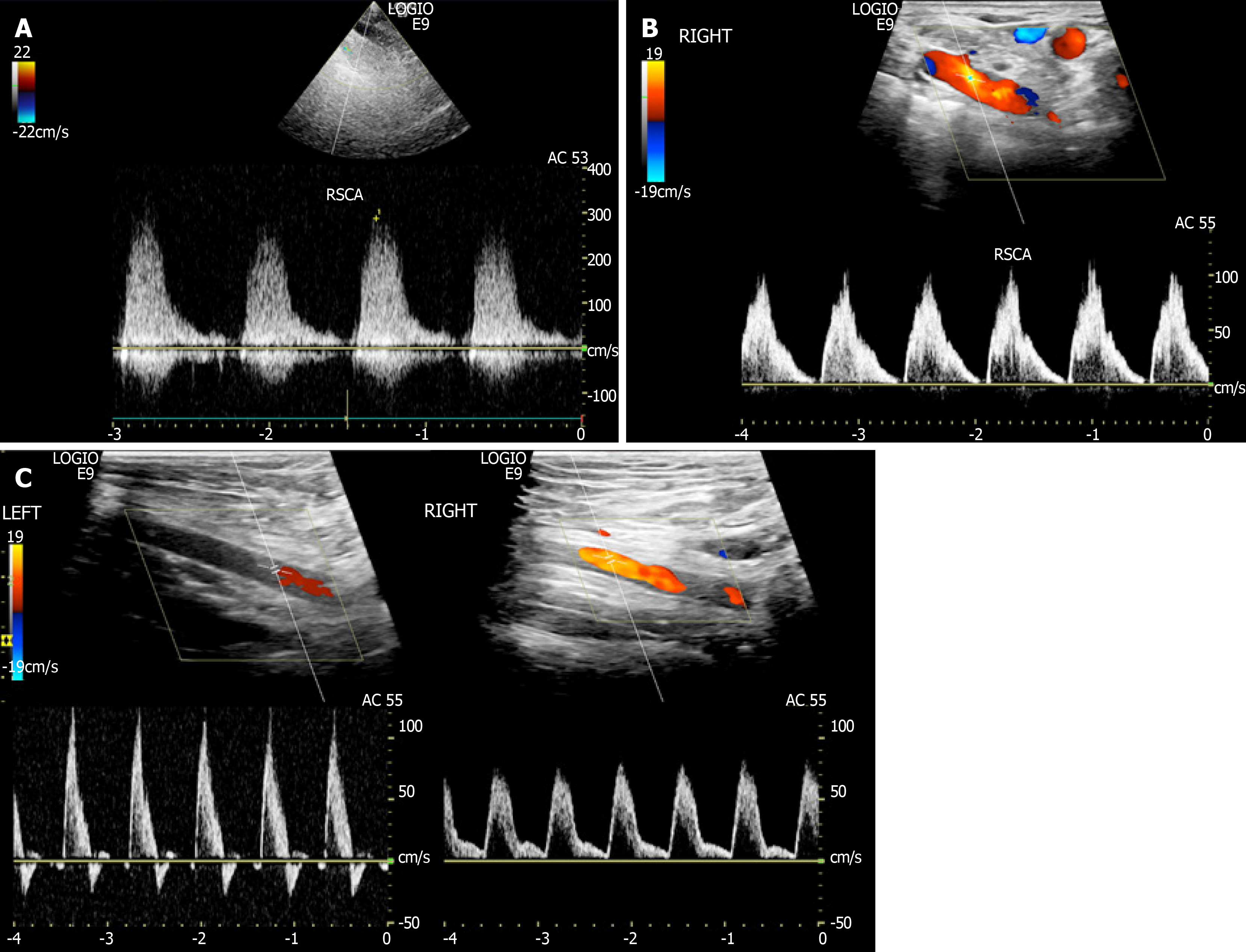
Figure 2 Ultrasound imaging examinations.
A: Ultrasound showed that the blood flow velocity at the right subclavian artery stenosis was up to 289 cm/s; B: Reduced at the distal segment presenting a single-phase wave change; C: Compared with the healthy left side, the flow velocity of the right axillary artery was significantly reduced and showed unidirectional changes.
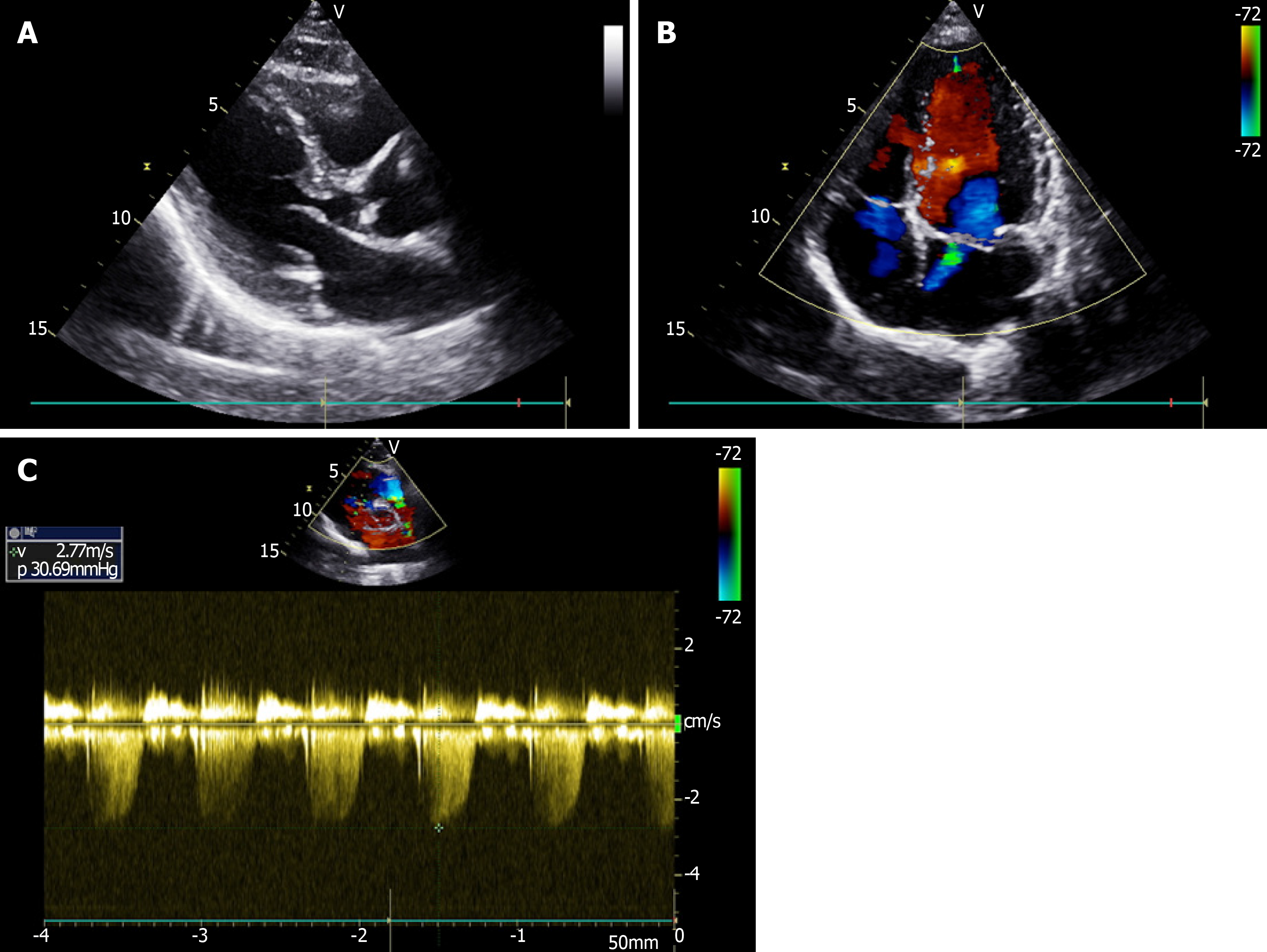
Figure 3 Echocardiography imaging examinations.
A: Showed calcification of the posterior mitral valve ring; B, C: Mild regurgitation of the mitral and tricuspid valves.
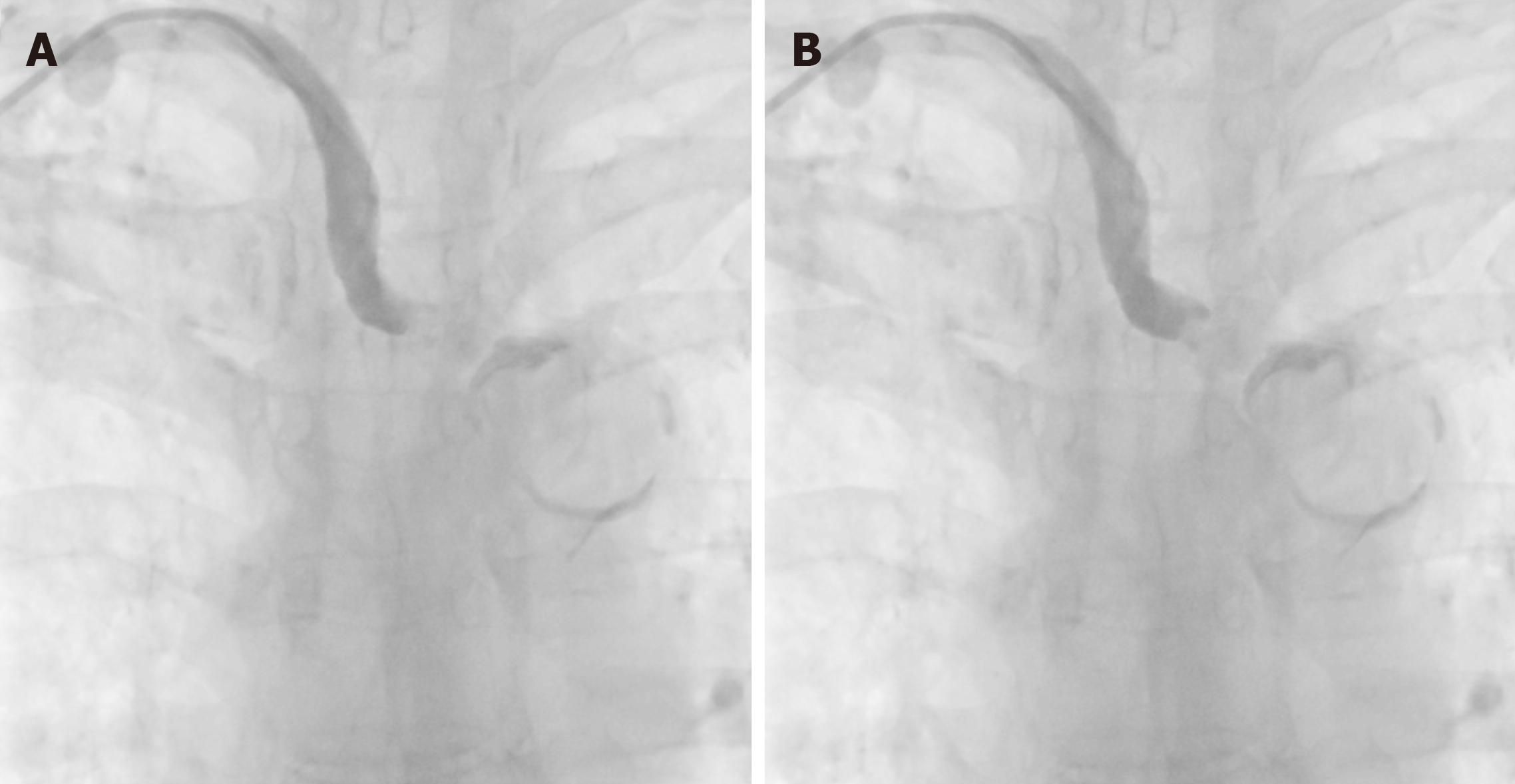
Figure 4 Digital subtraction angiography.
A, B: After puncture of the right radial artery, the proximal segment of the right subclavian artery showed no contrast medium passing through, indicating a broken end.
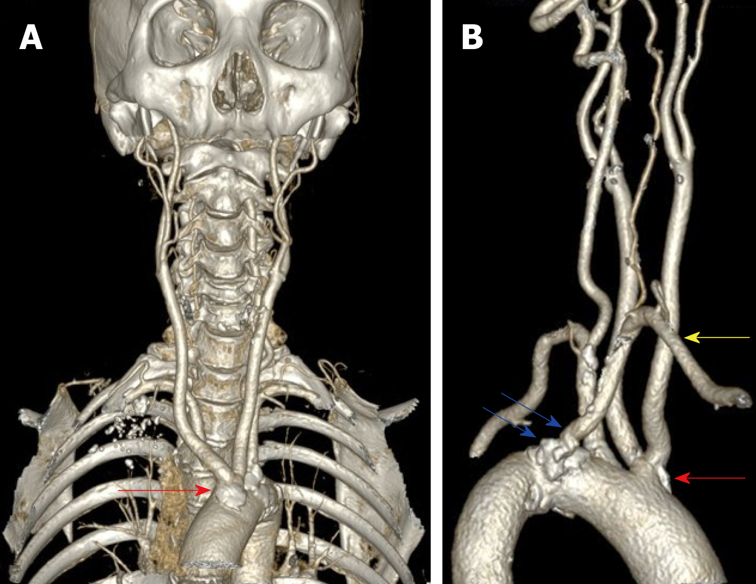
Figure 5 Computed tomography angiography.
A: Showed that the common trunk of the bilateral common carotid artery arose from the aortic arch (red arrow); B: The aberrant right subclavian artery (yellow arrow). Many calcified plaques were found at the beginning of the aberrant right subclavian artery and the common trunk of the bilateral common carotid artery (blue arrow).
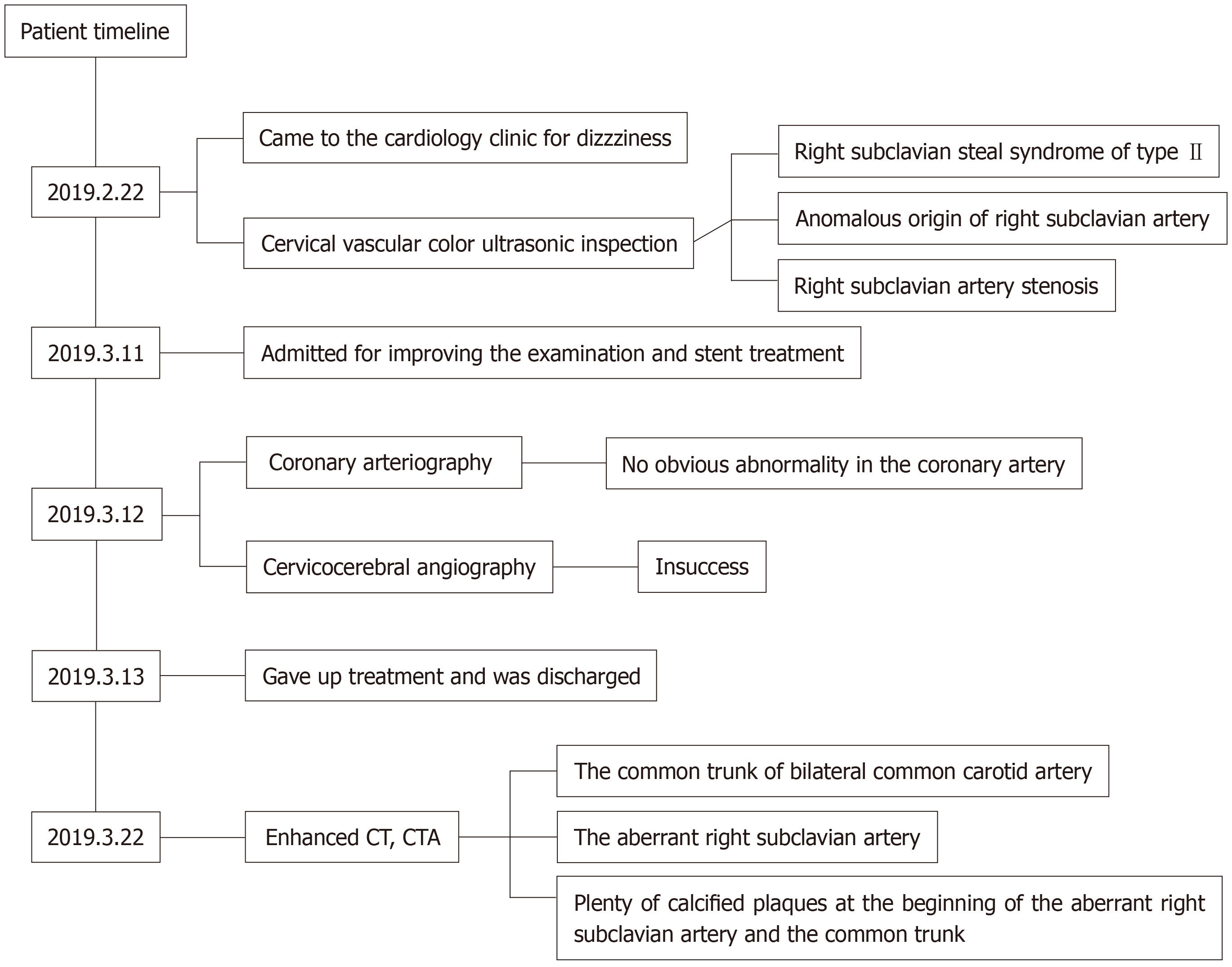
Figure 6 Timeline of patient diagnosis, treatment and follow-up.
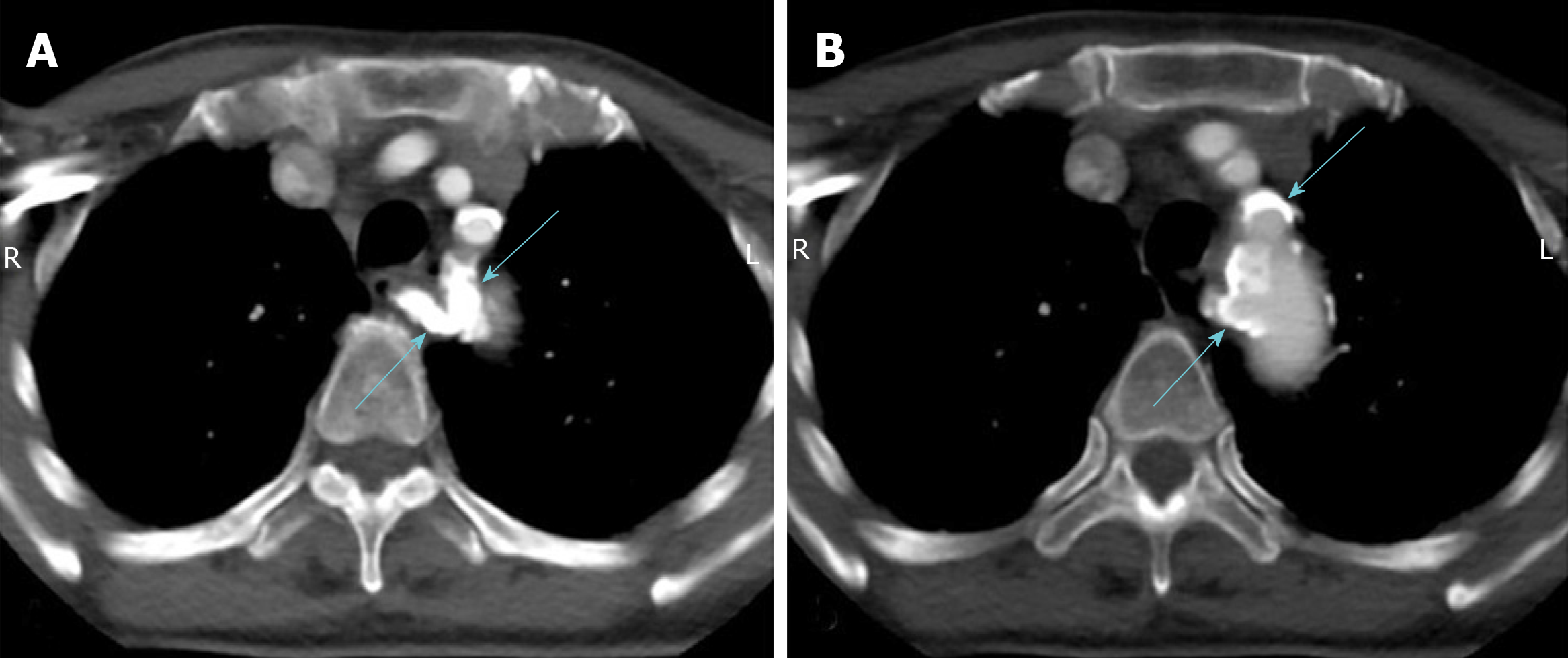
Figure 7 Enhanced computed tomography.
A, B: Most of the artery wall calcification plaques were concentrated at the beginning of the aortic branch artery, especially the aberrant right subclavian artery (blue arrow), which led to the aberrant right subclavian steal syndrome.
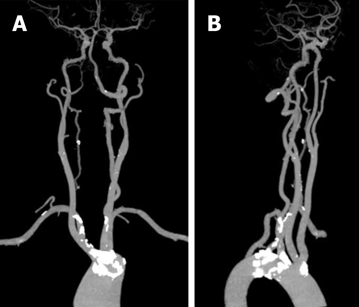
Figure 8 Computed tomography angiography.
A, B: Compared to the plaques at the aortic branches, most cervical and cerebral arteries had no stenosis in addition to the presence of scattered small calcified plaques.
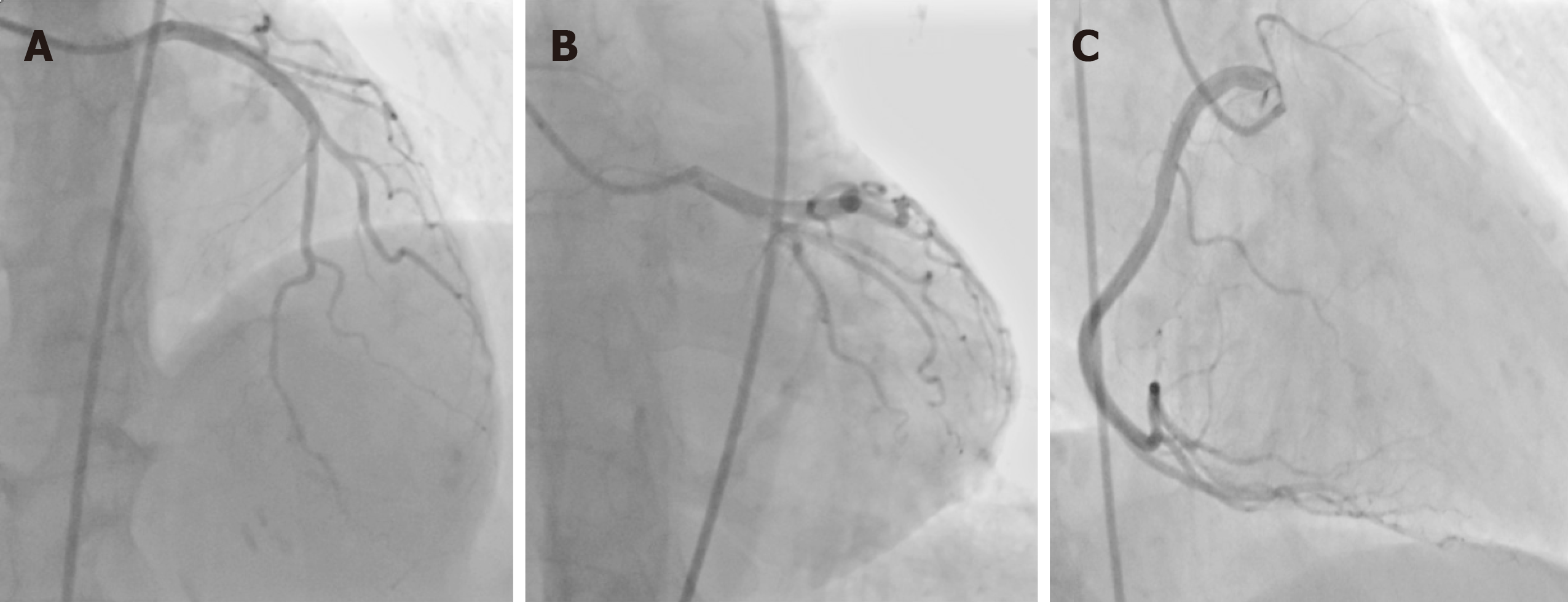
Figure 9 Coronary angiography examinations.
A, B: Showed no obvious stenosis of the left main, left anterior descending or left circumflex; C: Right coronary arteries.
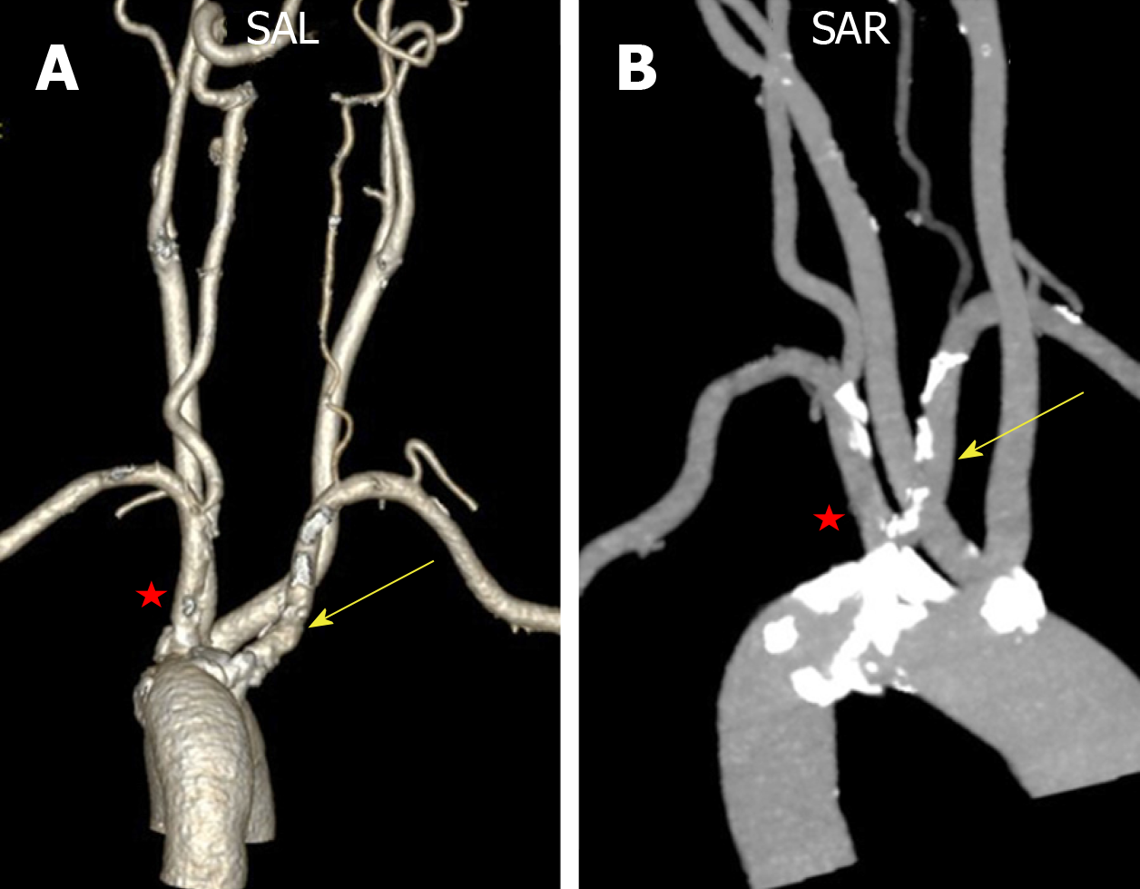
Figure 10 Computed tomography angiography.
A, B: Compared with the severe stenosis of the aberrant right subclavian artery (yellow arrow), the wall of the left subclavian artery was normal without stenosis (red star), although both had similar shapes and origins.
- Citation: Sun YY, Zhang GM, Zhang YB, Du X, Su ML. Bilateral common carotid artery common trunk with aberrant right subclavian artery combined with right subclavian steal syndrome: A case report. World J Clin Cases 2019; 7(21): 3639-3648
- URL: https://www.wjgnet.com/2307-8960/full/v7/i21/3639.htm
- DOI: https://dx.doi.org/10.12998/wjcc.v7.i21.3639


