Copyright
©The Author(s) 2019.
World J Clin Cases. Sep 6, 2019; 7(17): 2513-2518
Published online Sep 6, 2019. doi: 10.12998/wjcc.v7.i17.2513
Published online Sep 6, 2019. doi: 10.12998/wjcc.v7.i17.2513
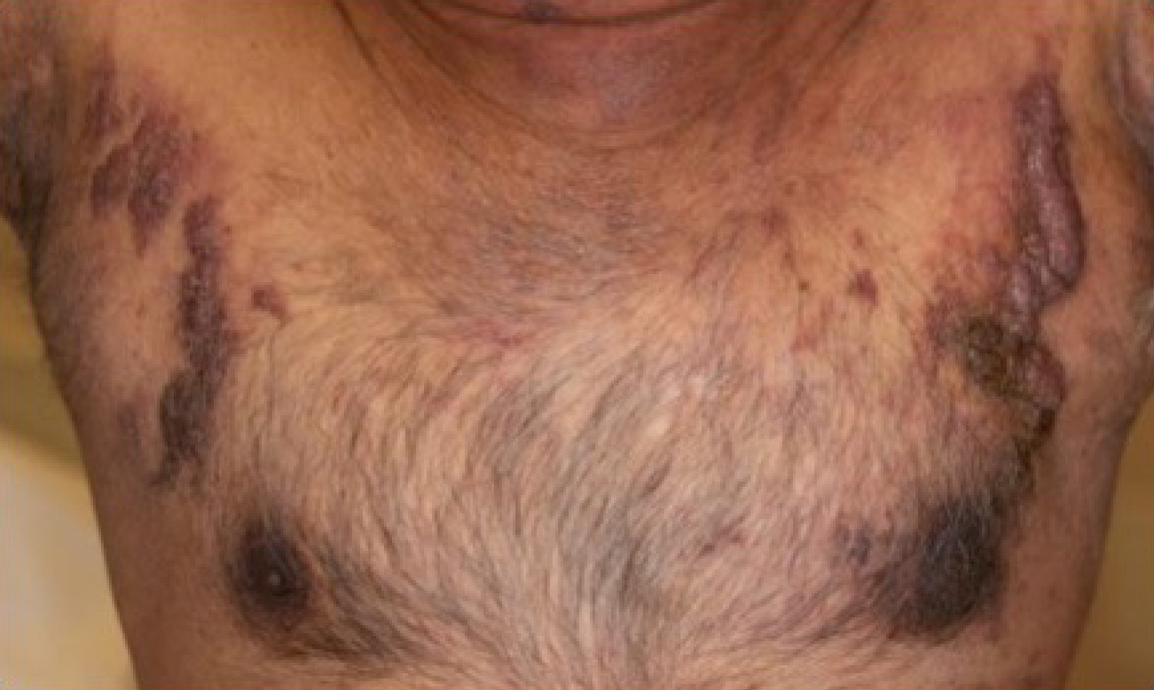
Figure 1 Nodular plaques involving bilateral pectoral districts.
Figure 2 Close-up of left (A) and right (B) chest skin lesions.
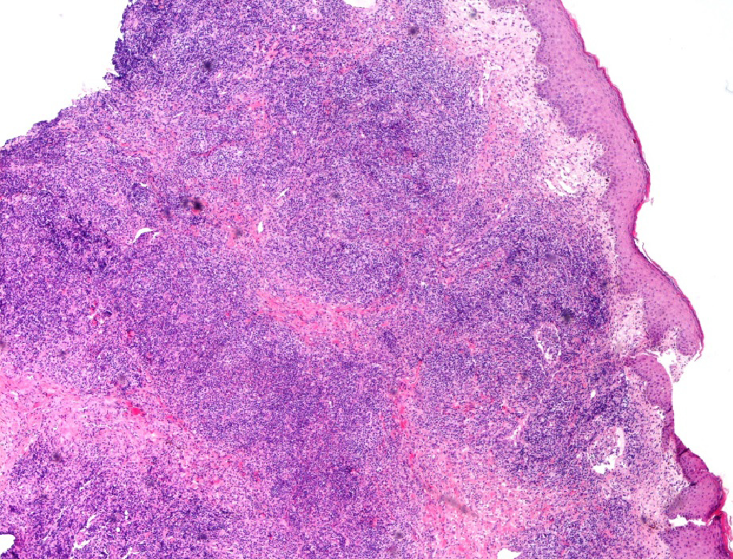
Figure 3 Dense dermic and hypodermic infiltration of lymphocytes, granulocytes (often eosinophils) and scattered Reed-Sternberg cells.
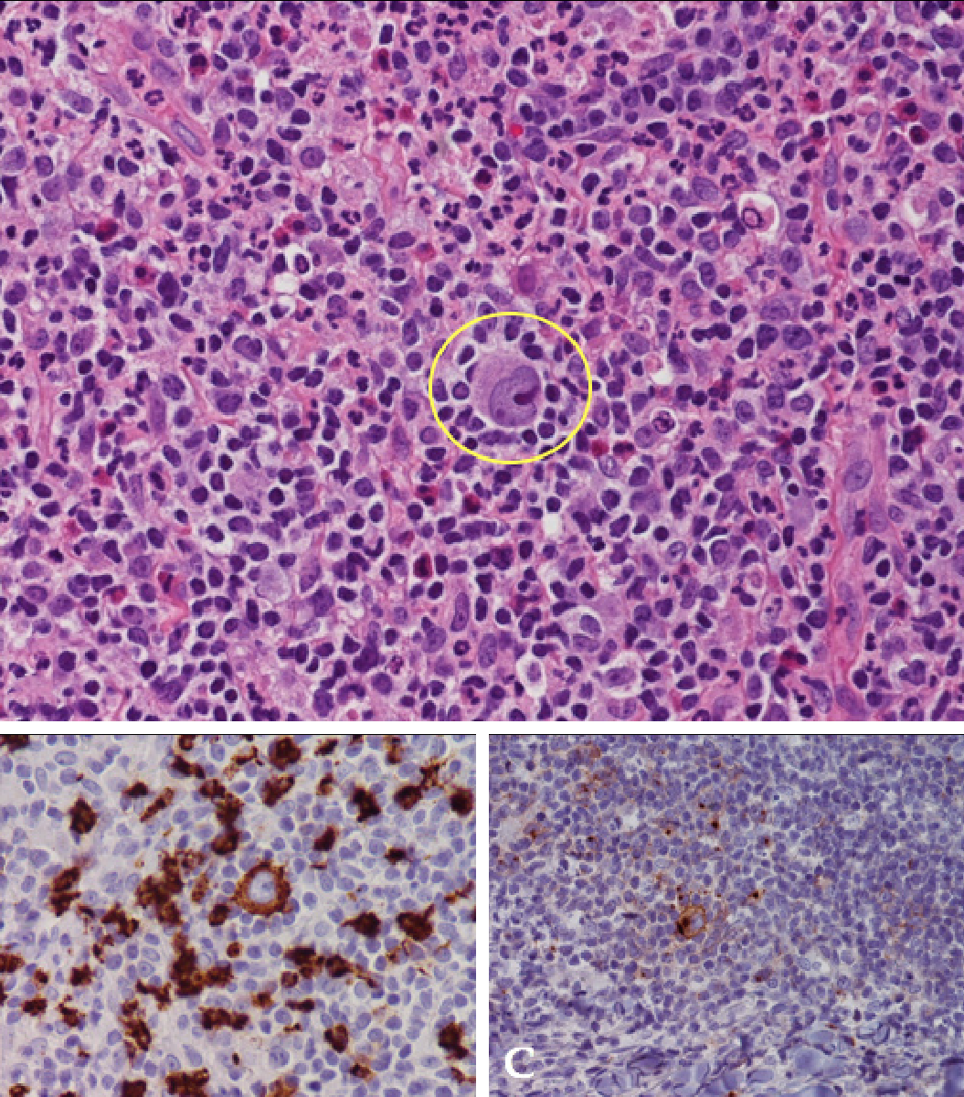
Figure 4 High power views highlighting a Reed-Sternberg large mononucleated cell (inset) in a background rich in eosinophils (A), CD30 (B) and CD15 (C) stains (HE 40 x).
- Citation: Massaro F, Ferrari A, Zendri E, Zanelli M, Merli F. Atypical cutaneous lesions in advanced-stage Hodgkin lymphoma: A case report. World J Clin Cases 2019; 7(17): 2513-2518
- URL: https://www.wjgnet.com/2307-8960/full/v7/i17/2513.htm
- DOI: https://dx.doi.org/10.12998/wjcc.v7.i17.2513


